
Earlier this year I picked up a rock with some orange lichen on it from a beach on the north west coast of Tasmania. I was interested to see how it looked under UV induced fluorescence and it did not disappoint (some initial fluorescence photos of it were shared here). A few weeks ago I came across an advert for someone who could make microscope slides of thin sections of rock (Dr Andrew Beard at Geology Hub) and I reached out to see if he would be able to make some thin sections of my Tasmanian rock sample so it could be viewed on the microscope. He was really helpful, and I sent him a few of my spare fused silica microscope slides (in case I wanted to try illuminating them with UV for fluorescence) and the rock sample and he made me a few slides to look at. Today’s post shared some initial images.
First a reminder, here’s the rock sample, front and back, in normal visible light and under UV induced fluorescence using 365nm light. It was about 3cm across and 1cm thick.

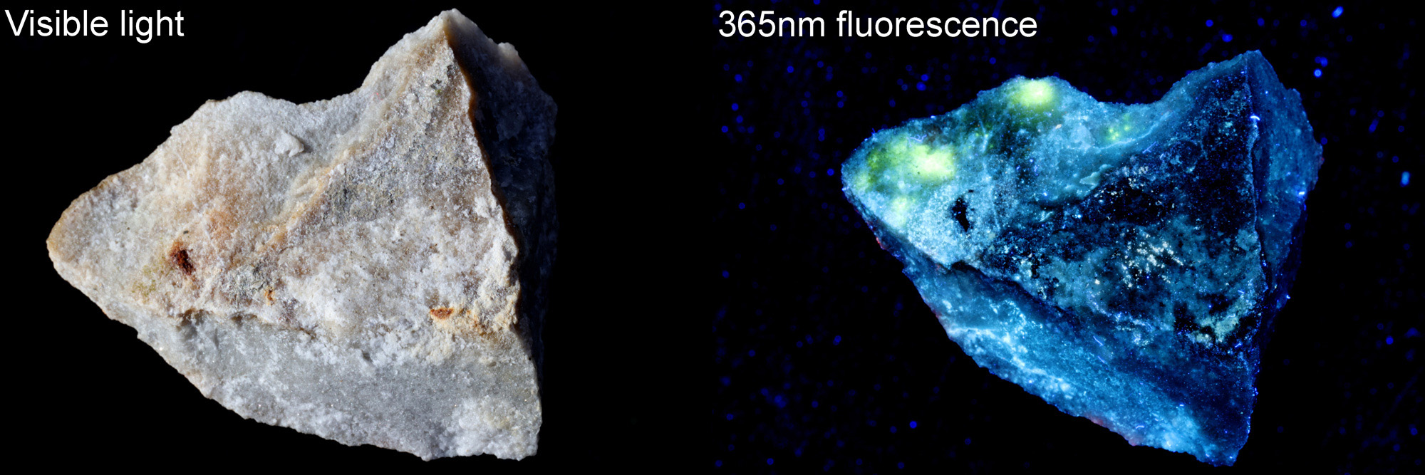
The front surface had an orange lichen on it it which also fluoresced orange under illumination with 365nm light. The rear had a couple of areas (towards the top left of the image) in which the matrix of the rock looked to be fluorescing yellow under 356nm light. I asked Dr Beard to prepare a few slides of the rock showing 2 specific areas – the orange lichen and the yellow rock matrix fluorescence. For the microscopy images, I used my modified Olympus BHB microscope, an Olympus Abbe condenser, a 10x Olympus UVFL NA 0.4 objective, an Olympus 2.5x NFK photoeyepiece and a Canon EOS R7 camera (auto white balance). Single images, no stacking, and full frames are shown without cropping.
Imaging of different slides done as follows:
Fluorescence: 365nm LED torch, with ZWB2 filter on the front (probably 2mm). 3mm thick GG420 filter on top of the photoeyepiece.
Bright field: White LED light. 3mm thick GG420 filter on top of the photoeyepiece.
Cross polarized: White LED light. Linear polarizer on the field lens. Moxtek linear polarizer on top of the photoeyepiece. Polarizers rotated to extinction.
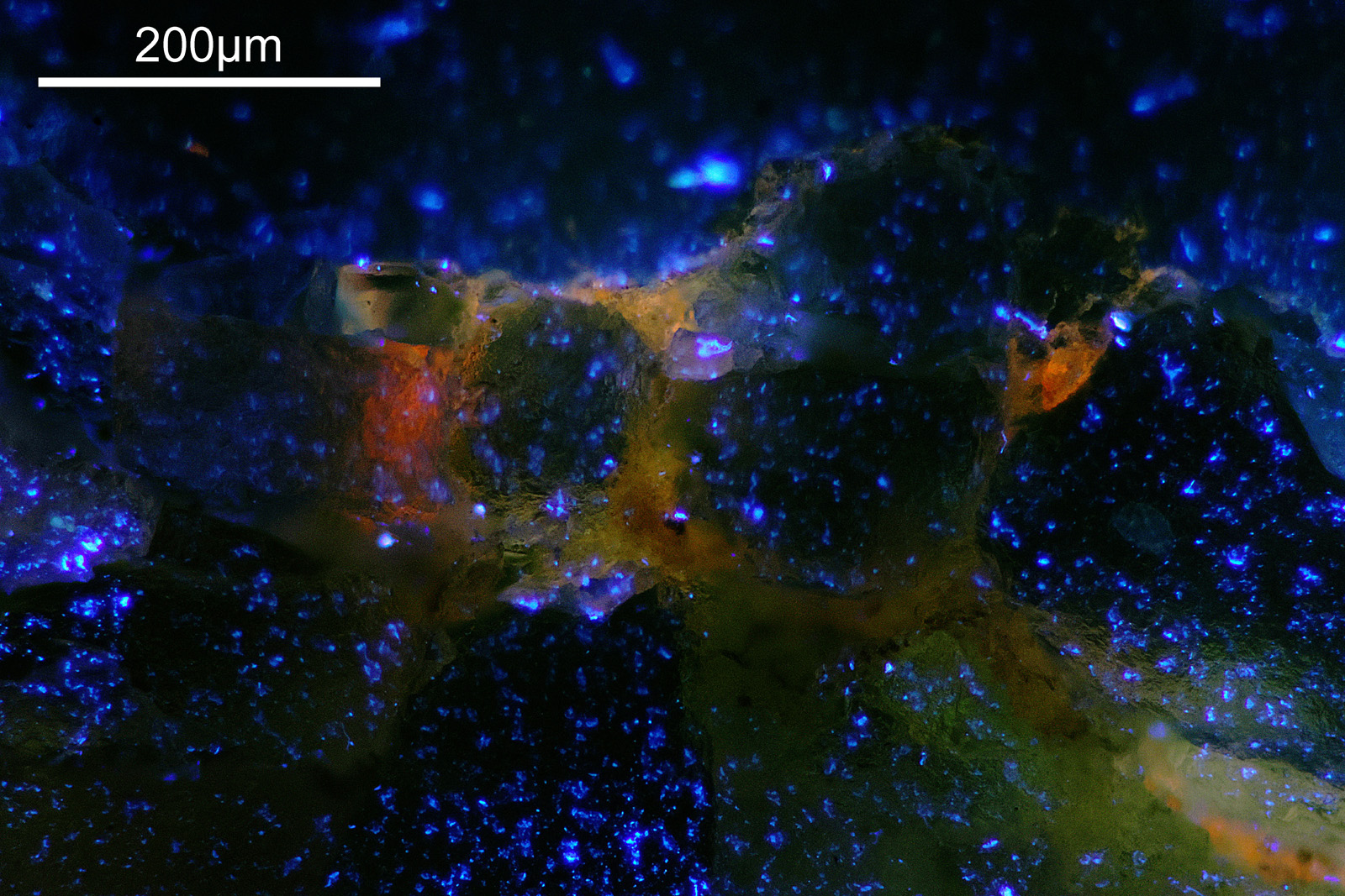
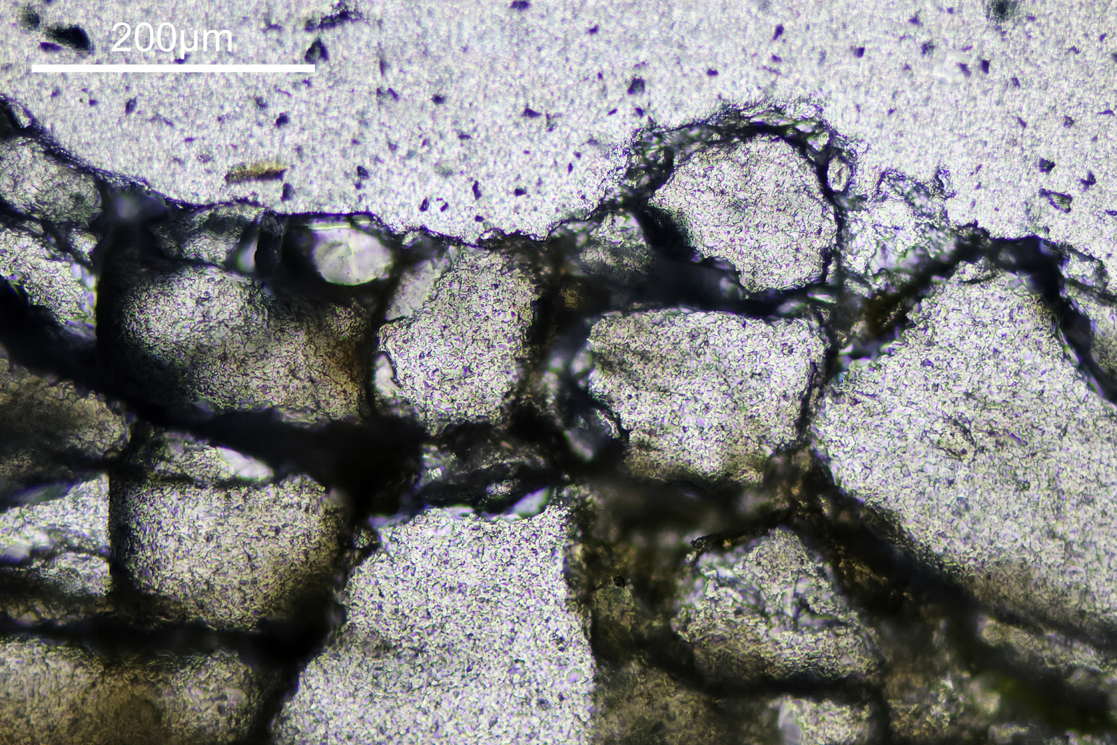
1st sample. A thin section (about 30µm) mounted in Petropoxy 154 resin and with a coverslip. The rock is at the bottom of the image, then a thin layer of lichen, and at the top of the image the mount. Shown as fluorescence, bright field, and cross polarized images.
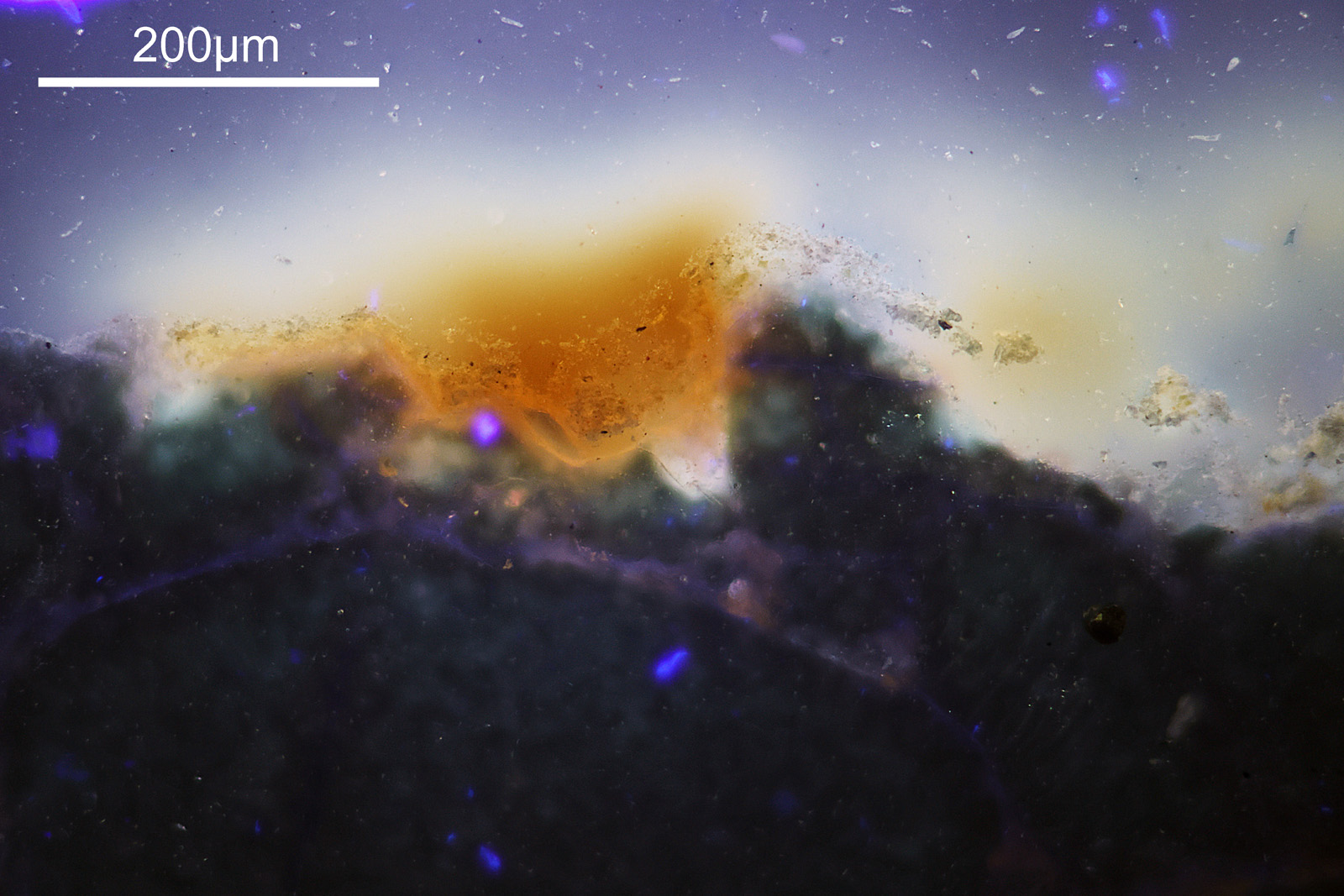
What’s going on with these images? In the bright field image, the thin rock sample looks fairly transparent as does the mount. The lichen looks to be quite dark in the bright field image, with a sort of ‘powdery’ appearance. The orange colour from the lichen seems to have dispersed into the mount a bit. This is also obvious in the fluorescence image where now the lichen itself looks to be quite transparent but there is a strong orange fluorescence surrounding it. In the cross polarized image the lichen is no longer really obvious (although there is a slight orange/yellow look to parts of the surface of the rock), but the different crystals in the rock are now really visible.
Given the bleed of the orange from the lichen in the Petropoxy 154 mount, Dr Beard suggested he could try using a different mount for some other slides, and so that is what he did.
Next, a thicker sample (about 60µm) with no coverslip.

Again the rock is towards the bottom of the image. The top is now open space. This sample looks different to the first as it is thicker and has no coverslip. It is therefore more 3D in appearance. The lichen now shows as a general orange glow at the surface of the rock in the openings in between the individual grains. Nice colours in the cross polarized image but as these are thicker and not well defined slices, it doesn’t look the same as the thin section in the 1st set of images.
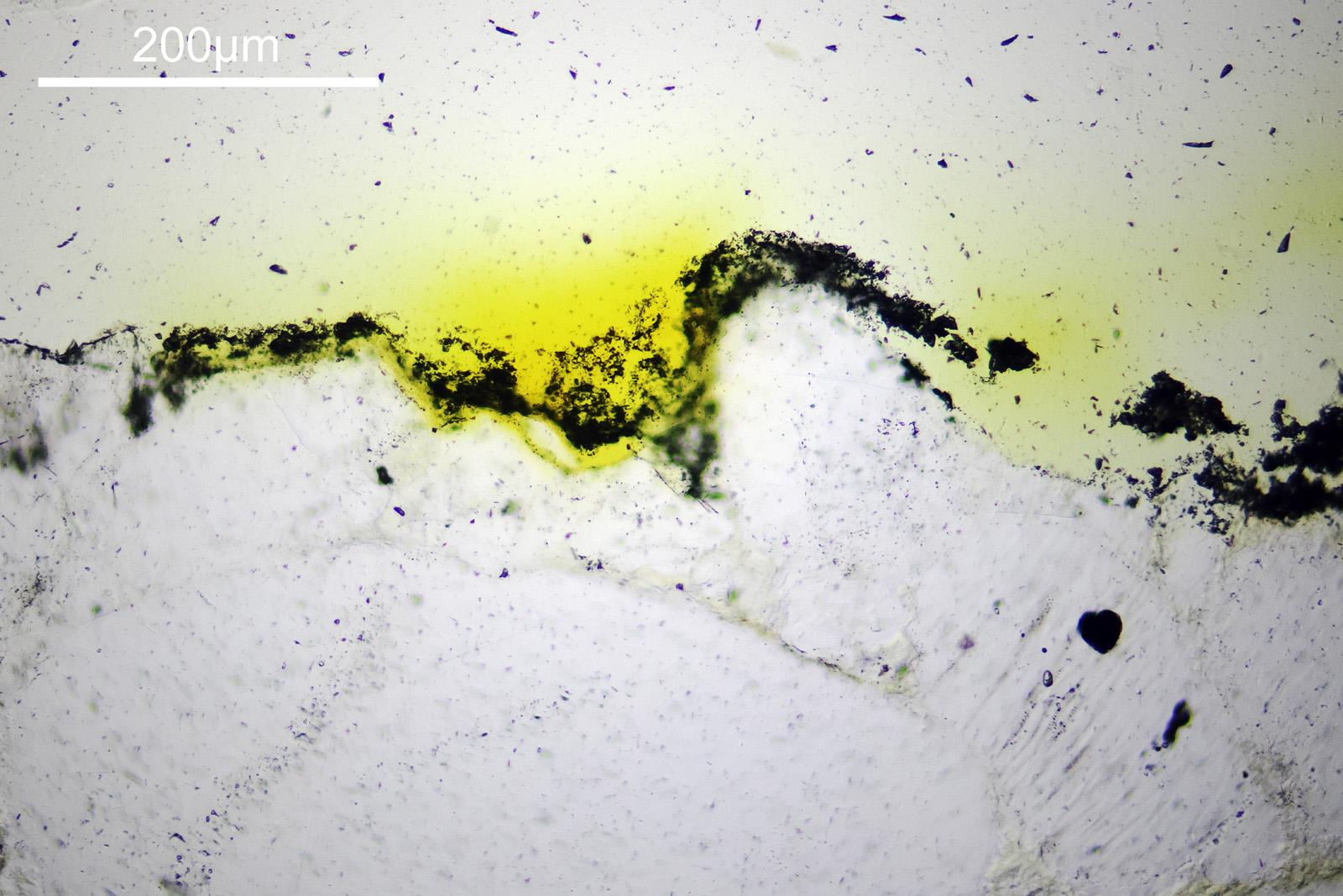
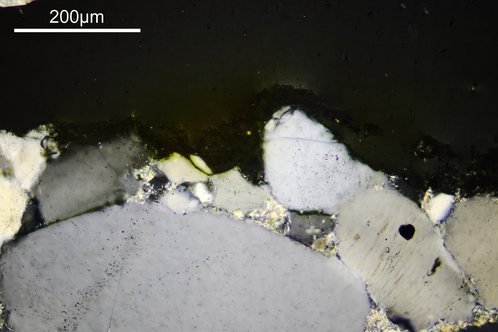
As a final set of images, these were also done with the newer mount, and as a thin section with a coverslip. This does not show the surface of the rock, but was an area of the matrix which had glowed yellow under 365nm light.
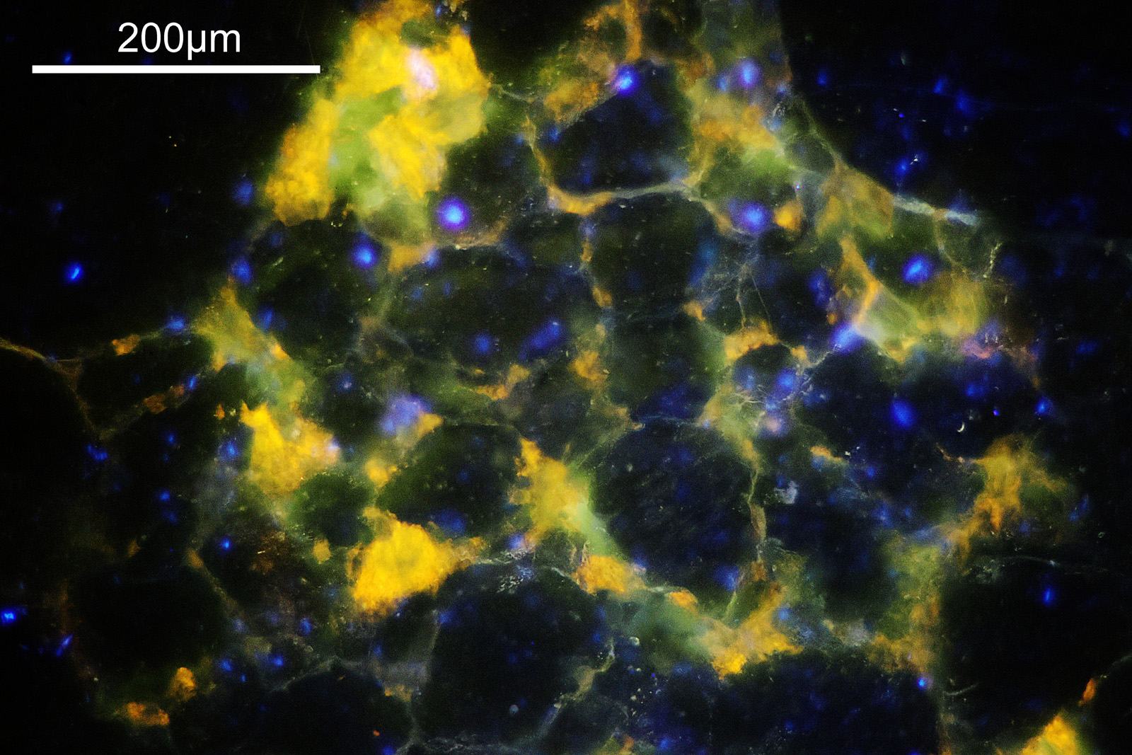
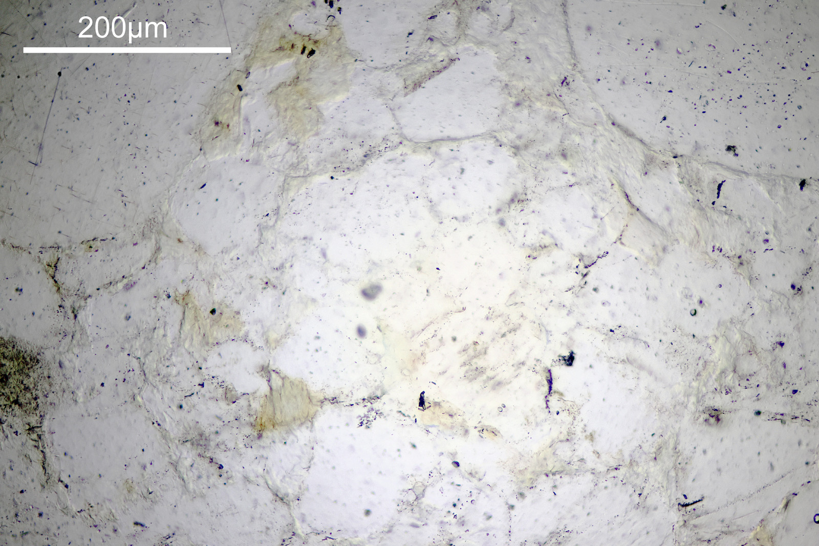
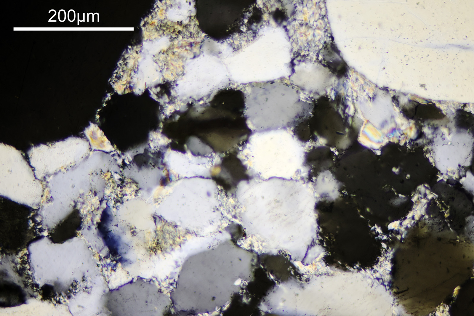
There is a strong yellow fluorescence which looks to be coming from whatever is in between some of the individual grains which make up the rock. This was quite heterogeneous across the sample, and this was an area where the yellow fluorescence was particularly strong. The bright field image doesn’t really show us a lot, although there is a slight colouration on the areas which fluoresce. The cross polarized image shows the typical appearance of a thin section again. The areas which fluoresce yellow look different – not black/grey/white but more rainbow coloured. Presumably these are a different mineral to the grains. It reminds me a bit of the Yooperlite mineral I imaged here. I used fused silica for these slides as glass will block short wavelength UV. It is a bit overkill for 365nm light, but does allow me to try shorter wavelengths in the future if I want to.
Overall I am really happy with the samples prepared by Dr Beard at Geology Hub, and they enabled me to have a look at the Tasmanian rock sample under the microscope. Changing the mode of imaging (fluorescence, bright field and cross polarized) had a huge impact on the appearance of the sample. As always thanks for reading, and if you’d like to know more about my work, I can be reached here.