
What you see when you observe the world is often a matter of perspective. Change your perspective and the world can appear a very different place. In our normal lives we view the world in the visible spectrum. What happens when we change that? If we polarise the light? If we look in the Ultraviolet? If we do both? How about fluorescence? These different approaches can allow us to see things which are often hard to observe using our normal approach.
Skin photography is used widely to image skin and scars, for cosmetic, pharmaceutical and forensic applications. When I was a young boy, I did many reckless things, and like many young boys I got a good selection of scars to remind me of them. One is a scar on my index finger on my left hand – a linear scar about 2cm long with 2 stitch marks running across it. I’ve had this for over 30 years now and it has faded a lot which make it hard to image. I got to wondering whether by changing how I imaged my hand, I could increase the contrast of the scar and make it easier to observe. So I imaged my hand with a number of approaches;
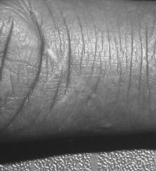
Normal visible light.
Cross polarised visible light.
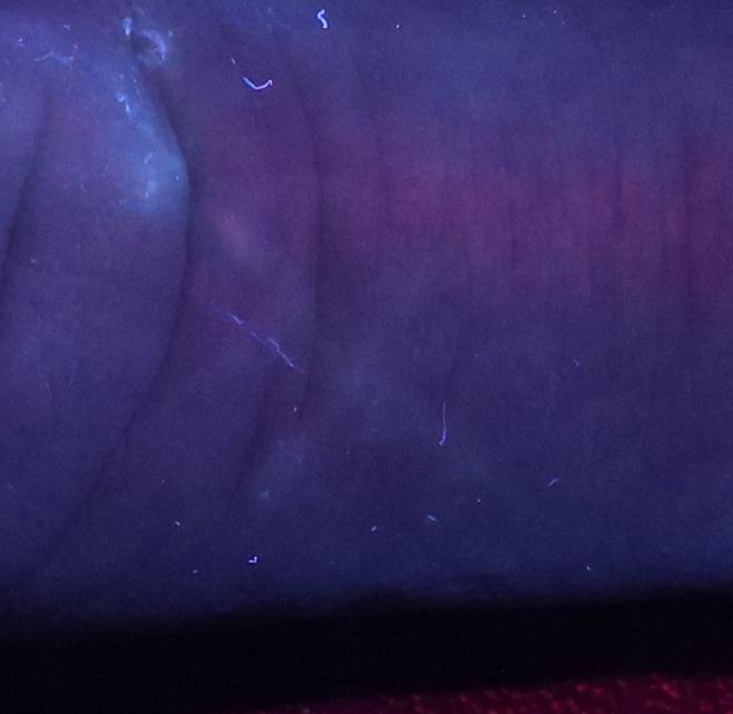
UV induced visible light fluorescence.
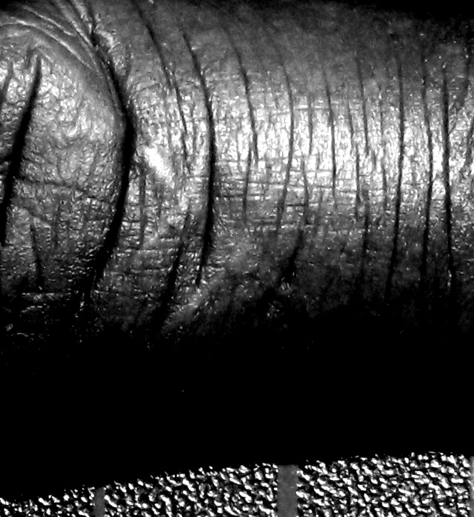
Reflected UV light.
Cross polarised reflected UV light.
The images from these are shown below.
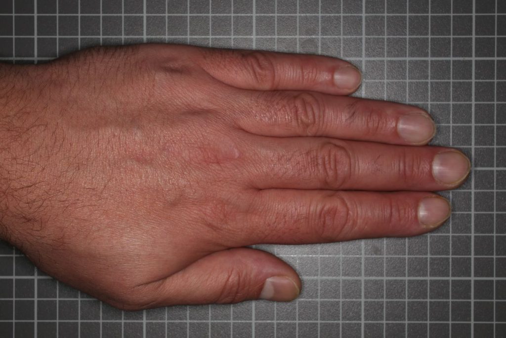
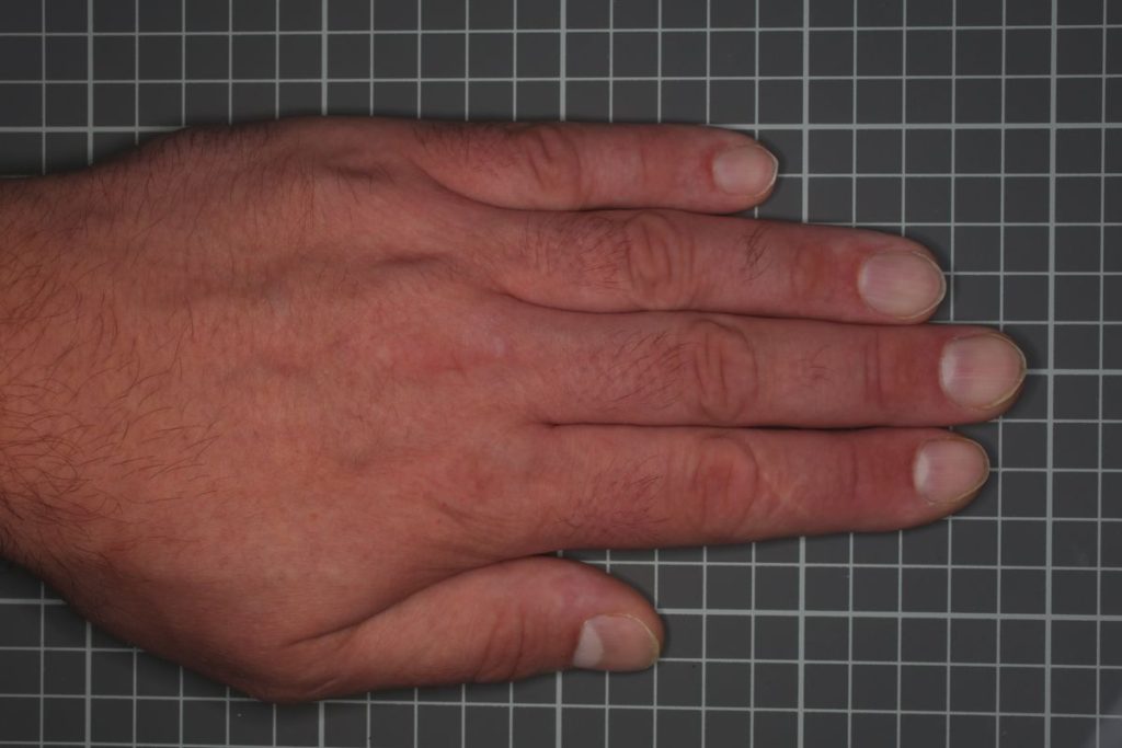
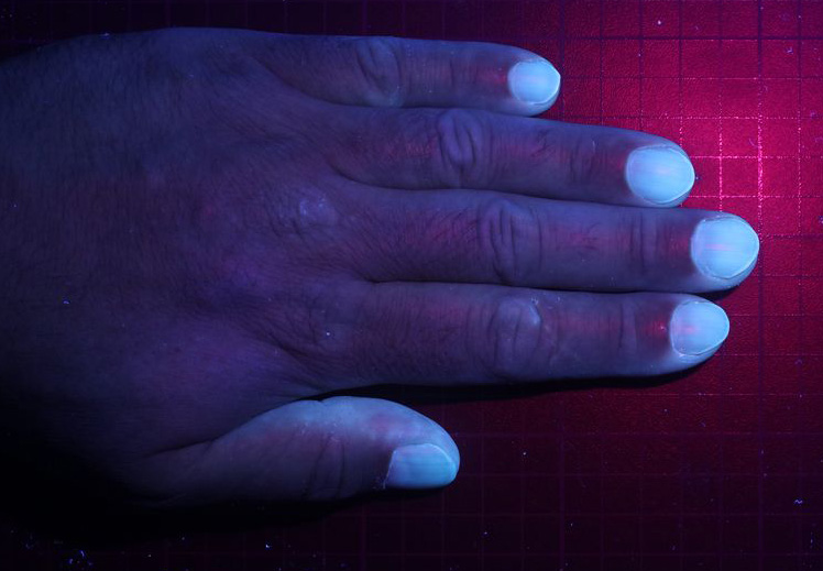
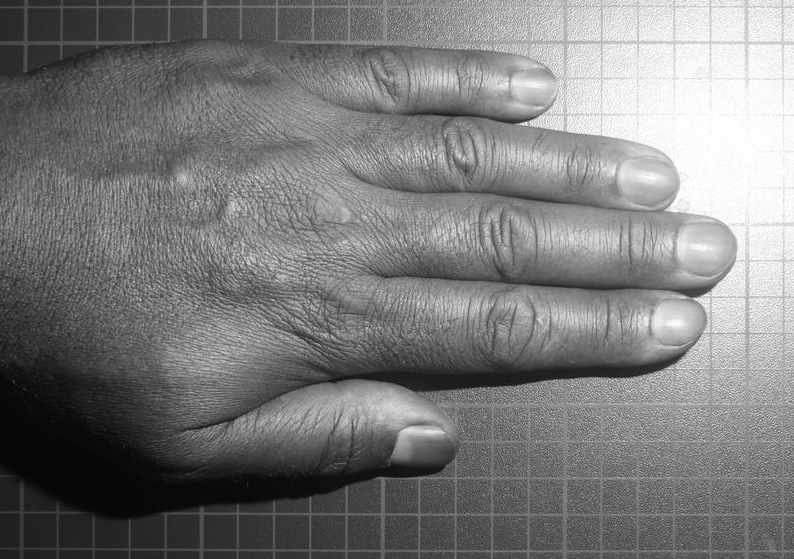
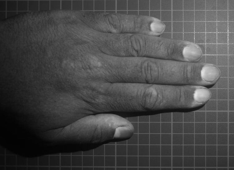
As can be seen the lighting and imaging has a huge impact on the appearance of the skin. The forefinger scar can be seen, but barely, and it is more obvious on the cross polarised visible image (where the shine on the skin is removed, and the paler scar is easier to see), and the UV reflectance image (where the surface texture is actually emphasized).
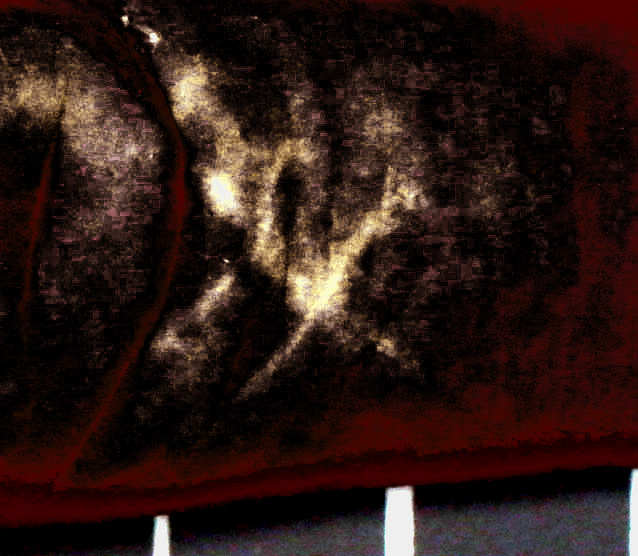
These is complex images to observe – there’s a lot of features such a wrinkles and lines, contrast changes, and even colour changes. This would make it hard to try and automate detection of the scar in amongst everything else. How about when the images are cropped to show the area of the scar, does that make it easier to see?
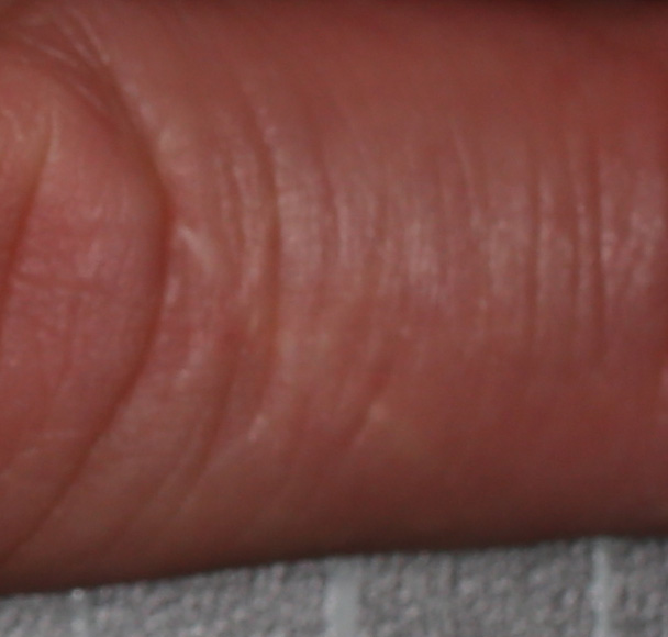
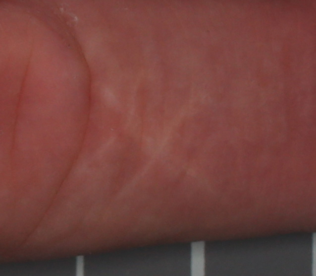
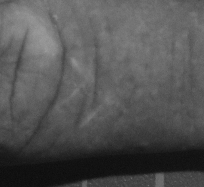
Even now, cropping the images to look at the scar it is hard to visualise, although it is easier to see in the cross polarised visible, and standard UV reflectance images. These images can then be taken further, using modifications such as brightness and saturation boosting, selective colour replacement, and inversion to increase the visibility of the scar.
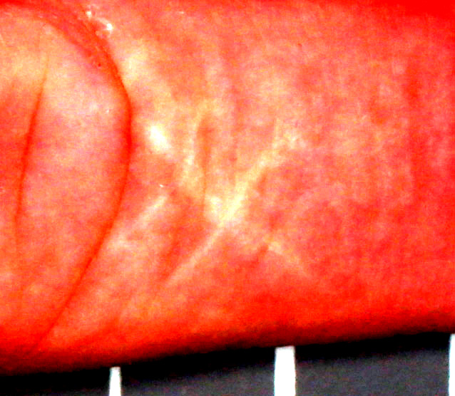
A simple brightness and contrast boost makes the scar more visible, and would help with identifying it in an image. How about taking it further? Further work on the visible cross polarised image gives using selective colour replacement gives this.
Through a selective colour swap of the cross polarised visible image, the scar can be made to stand out against the surrounding skin – the linear feature and stitch marks becoming easier to observe.
Very often in research we are looking for very subtle features set against a complex background, and a simple photo just isn’t enough. Changing the wavelength of light used, adding cross polarisation, and performing image modification can all be used to boost contrast between the feature we are looking for and the background, making the invisible, visible again. It’s just a matter of perspective…..
If you’d like to know more about my imaging work, please contact me here.