
Anyone who has chatted to me will probably have gathered that I have a huge soft spot for Tasmania, as it doesn’t normally take me long to start talking about how amazing it is. In previous visits I have taken my UV camera along to capture some photos there (UV levels are very high in Tasmania which make UV photography easier, but has a nasty habit of burning you if you forget your sunscreen and hat). I’ll share some of the images from these visits in the future as they demonstrate nicely the effects of UV imaging compared with visible light and IR imaging when photographing the landscape. To start things off though, the first post on this topic is something a little different – UV induced fluorescence photography. This uses UV light to illuminate the subject, and the visible light this produces due to fluorescence is then photographed. The subject for this experience was a rock with some of the orange lichen on it. Orange lichen is present across most of Tasmania and makes for striking landscape photos, and it got me wondered what this material looked like under UV induced fluorescence. I am certainly not a lichen expert, so this will concentrate more on the imaging and less of what specifically the lichen is.
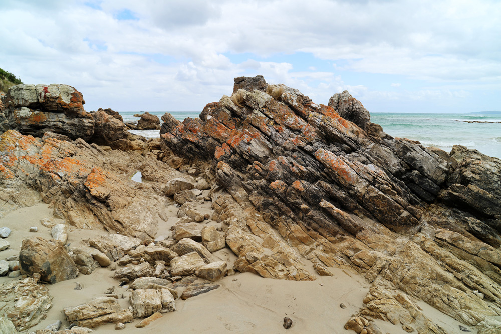
The rock itself came from Green Point Beach in the North West of Tasmania (part of the Rocky Cape Group). The rocks on the beach had some damage from regular strong waves, so I picked up a piece a few centimeters across which looked to have recently broken off from the main outcrop. The rocks in this part of Tasmania are old. Very old. They are about a billion years old, and appear to be something like quartz arenite.
Here’s the front of the rock in normal visible light and in UV (365nm LED light) induced visible light fluorescence.

And the rear of the rock, again in visible light and UV induced fluorescence.
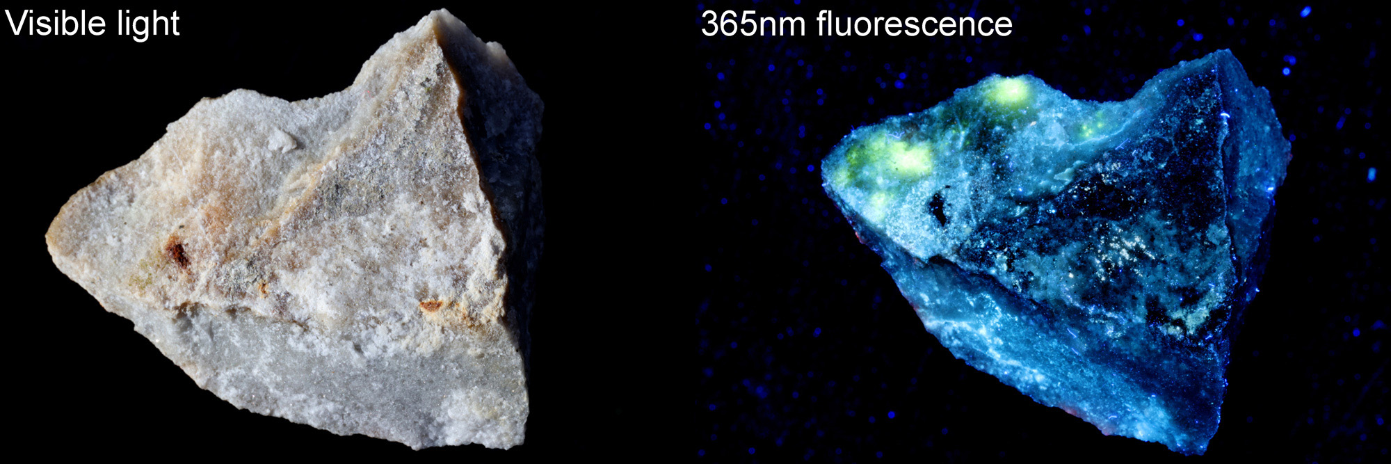
The front surface of the rock sample shows the vivid orange colour in visible light. Interesting this lichen also fluoresces orange under illumination by UV light. The matrix of the block itself mainly fluoresces blue, however there are some bright yellow regions as well, perhaps indicating slightly different mineralogy in those areas.
Here’s the rock formation on the beach.
This does raise an interesting possibility – could UV induced fluorescence be used in the image of lichens to help with their identification? Do all lichens which are orange in the visible spectrum fluoresce the same way? This is something I don’t not know, but UV fluorescence is used in many research areas so might be worth trying in the future. It would also make for an interesting form of landscape photography – light painting the scenery with a portable UV light and imaging at night or in twilight.
Technical details. For the fluorescence image the light source was a 365nm LED light which was filtered to remove visible light. the camera was a Canon Eos R7 and a 420nm long pass blocking filter was use to eliminate any reflected UV light. A daylight white balance was used for the UV induced fluorescence image. The lens was a 105mm Rayfact (UV Nikkor) macro lens at f22. ISO 200 was used for the images, and the UV fluorescence image was of course taken in the absence of natural visible light. A black background and sunlight was used for the visible light image.
As always, thanks for reading and if you’d like to know more about this or any other aspect of my work, I can be reached here.