
Testing, testing, testing. As always with new equipment and methods it is helpful to test them to see what they are capable of and where their limitations are. With my UV microscope, I recently had a test slide made using quartz components and which had a variety of diatoms mounted on it. These diatoms have very small features (often sub micron in size) and are great for testing optical setups. Also, being made from quartz it is usable in the UV where glass would block the light.
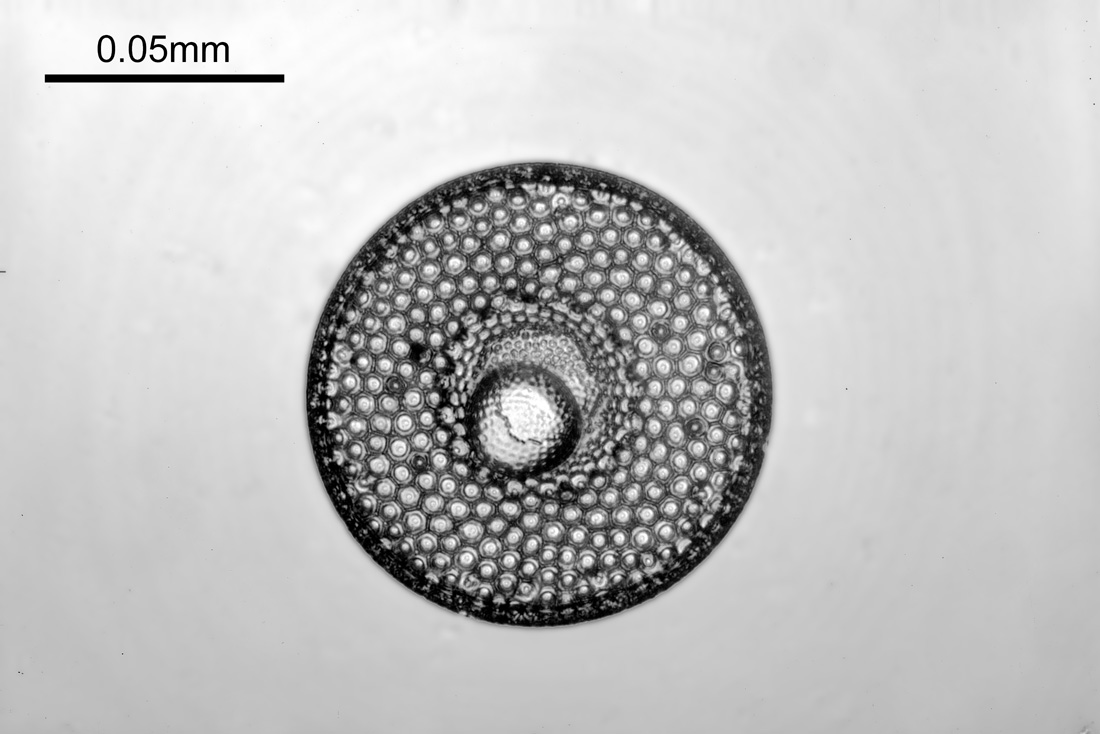
Todays images were done with 3 different objectives – but before we get into the specifics of the objectives, I’m going to share one of the images produced, because, it is pretty special. This was made with the Leitz 40x NA 0.65 objective which will be discussed below and is a 313nm brightfield transmission image.

Back to the objectives. Three were used for this – a Zeiss 32x NA 0.4 Ultrafluar, a Leitz 40x NA 0.65 UV objective, and a 36x NA 0.5 Beck reflecting objective, all of which are shown below.
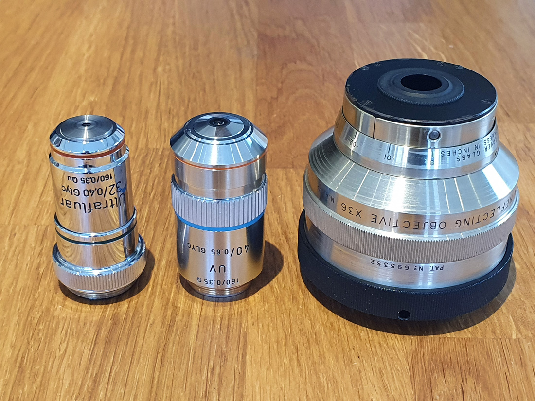
I normally use the Ultrafluar when I need an objective in this sort of magnification range, but wanted to check the other two out. The Zeiss and Leitz were both used with glycerine immersion fluid, and the Beck was used dry. The Beck is very different to the other two being a mirror lens, and I checked the alignment of it before using it. The diatom was imaged at 313nm, and each image is a stack of about 13 images made in Zerene stacker. Some processing of the final images was done in Photoshop, but all images were treated similarly. They have been reduced in size for sharing online.
First the 32x Ultrafluar.
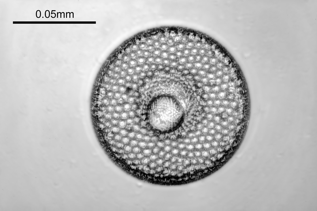
Next with the 36x Beck reflecting objective.
And finally with the 40x NA 0.65 Leitz UV objective.
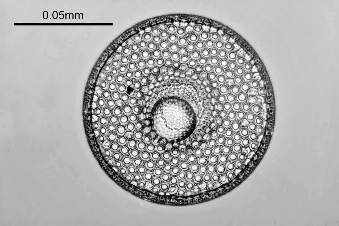
Obviously the field of view is different with the three objectives as their magnifications are not the same. The 32x Ultrafluar image is certainly very good, especially for a NA 0.4 objective. The 36x Beck reflecting objective image is lower contrast and not as sharp, but still shows a lot of detail. However the star of the show here was the Leitz 40x NA 0.65. The final image it produced was extremely sharp and full of detail.
Not much is known about the Leitz 40x UV objective, certainly nowhere near as much as about the Ultrafluar objectives, however I have written a bit about it here. To try and get a bit more detail on the resolution it was producing I did a crop of the original image in ImageJ and took some measurements. The area assessed is shown below.
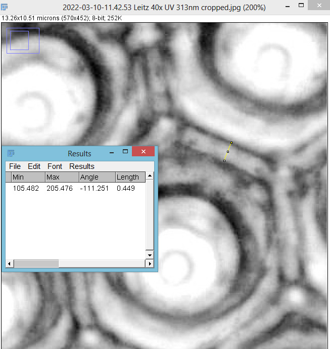
The yellow line in the image above is 449nm long, so it looks like this objective is easily resolving features below 0.5micron in size. Abbe’s resolution equation says that this objective should have a resolution of 241nm when used with 313nm light, and from the looks of this image it probably isn’t far off that. Makes me wonder what my Leitz 100x NA 1.20 objective is capable of, but that will have to wait for another day….
I’m certainly no diatom expert, but they are beautiful structures and make for great photos, so I will be doing more in the future. It’s been great to finally be able to test some of my UV objectives with samples like this, and to be able to produce some real life images with these historic optical pieces. Thanks for reading, and if you’d like to know more about this or any other aspect of my work, I can be reached here.