
A short update today. I returned a diatom slide I have imaged before – the Amphipleura linheimerii slide originally imaged here. I wanted to try imaging it at 365nm, but using a Watson Quartz cassegrain condenser that I have on loan (a very special condenser, which I will try and write more about in the future). In summary, the condenser is a high NA dark field condenser, and being made from quartz has great UV transmission. This post shares an image from the slide using 365nm dark field illumination.
First though a quick note about the setup. Microscope was my UV modified Olympus BHB. Light source was a 365nm torch slid into the light port of the microscope. Condenser was a Watson quartz cassegrain one, which was used with glycerine/water as the immersion fluid. The objective was an Olympus 40x NA 1.3 silicone immersion lens, which I used with glycerine/water as the immersion fluid. Photoeyepiece was an Olympus 2.5 NFK. Then I used 2 stacked Edmund Optics 365nm, 10nm FWHM bandpass filters. Finally a monochrome converted Nikon d800 which is UV sensitive.
Ok, the image – this has been cropped slightly, and has been reduced in resolution for sharing.
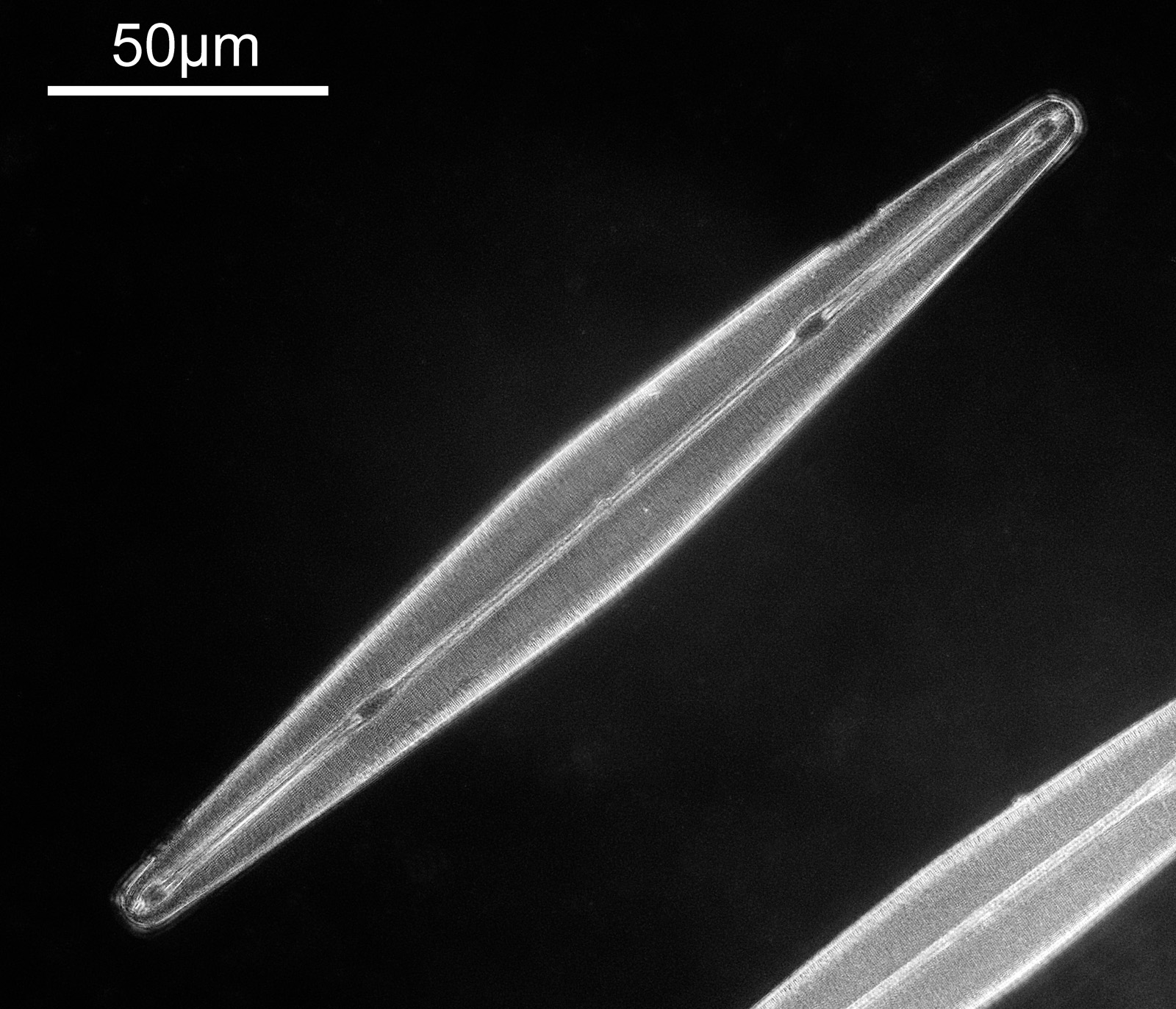
There are two examples of the diatom on the slide, and part of the other one can be seen in the lower left of the image above. There is plenty of detail in the image, but perhaps that does not show up as well with the lower resolution image being shared here. Here’s a crop, at original pixel resolution.
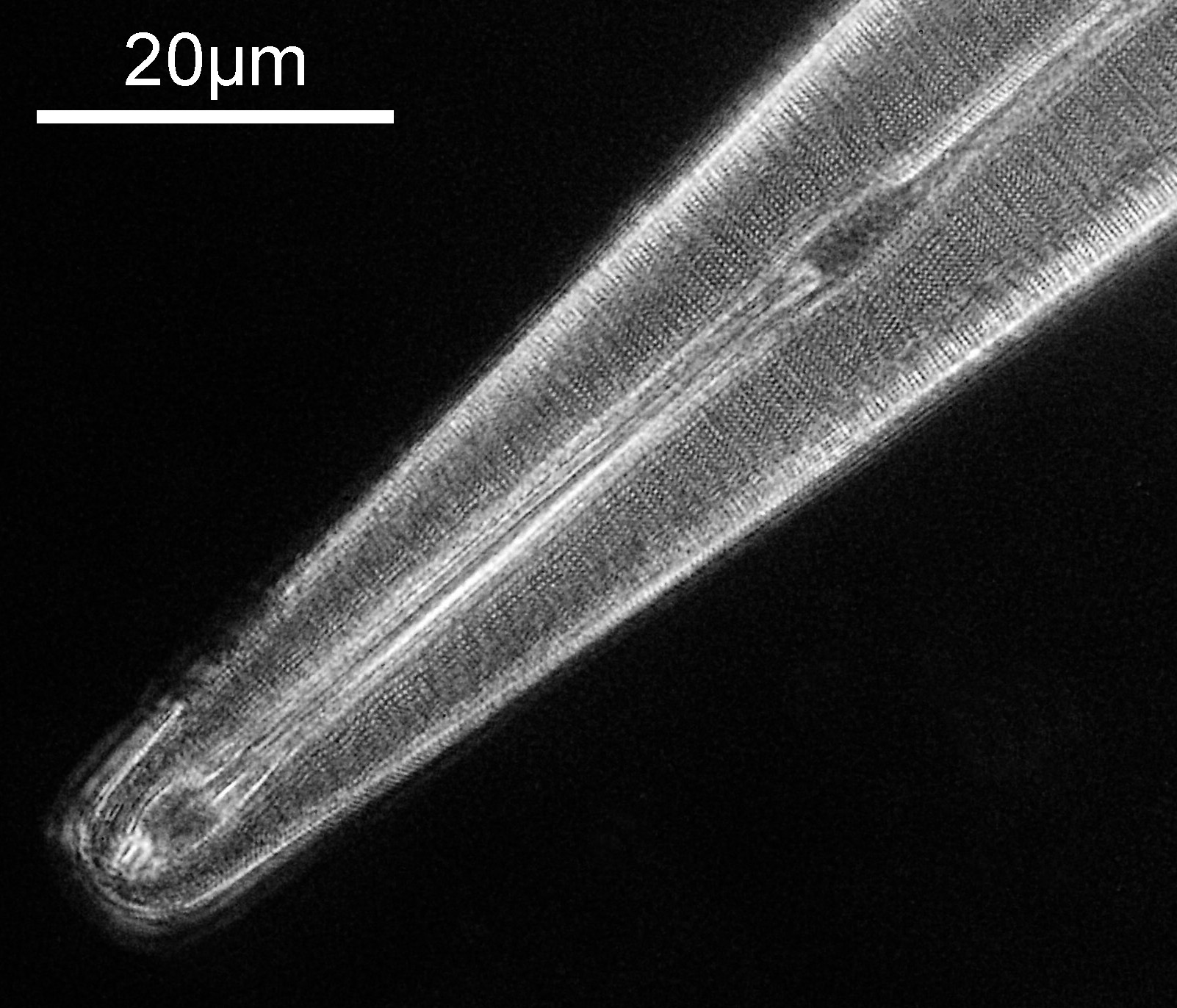
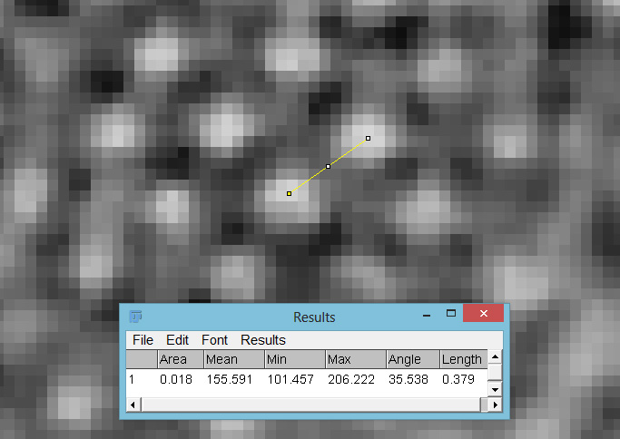
With the cropped image some of the smaller features of the diatom now become visible. The distance between the small white dots was measured on using ImageJ was was found to be about 380nm (length 0.379 in the image below, as it was calibrated in microns).
A few things to note with this setup. Glycerine/water was used as the immersion fluid, while the objective was designed for silicone. Glycerine/water was used for a few reasons – firstly, I had some, secondly, it is a similar refractive index to the silicone, and finally it is much, much easier to clean. However I have to acknowledge this may impact performance of the objective. Being quite a low magnification objective, the field of view was large. As such the resolution achievable by the objective is getting close to the actual pixel resolution of the camera. I did try taking RAW files and then converting them to monochrome in Monochrome2DNG to get more resolution, and I even tried frame averaging in the camera, but neither of these improved things noticeably and I just stuck with the original single frame jpg captured in the camera in the end. The slide itself was not very UV transparent. I suspect if I measure the transmission spectrum of the slide, it will not be great at 365nm. As such this was not the ideal subject. I am also not sure I got the iris on the objective fully optimized, so perhaps a little more resolution can be squeezed from this setup.
Diatom imaging in the UV continues to be a fascinating journey, and one i will continue to pursue. As always, thanks for reading, and if you’d like to know more about my work, I can be reached here.