
A bit of a fun experiment today. The images we create depend greatly on how we light our subject. Changing the lighting can have a drastic impact on the final appearance of the photo. This post shows how using a historic Watson Holoscopic condenser can be used to image diatoms, and how the picture look compared with straight brightfield, and oblique illumination.
The subject was a Darlaston slide with two Amphipleura lindheimeri diatoms present and a two circular diatoms as well. The microscope was my usual modified Olympus BHB. Objective was a 40x Olympus UVFL PL NA 1.3 silicone immersion one with an adjustable iris (the adjustable iris was important for this experiment, more on that later). I used 450nm filtered LED light, and images were captured on my monochrome converted Nikon d800 camera. For condensers I used an Olympus Aplanat Achromat one (normal brightfield and oblique brightfield) and the Watson Holoscopic condenser. For both of these I oiled them to the underside of the slide using immersion oil, and I also used that for the objective. The objective was designed for silicone oil, but I didn’t have any of that, so just used immersion oil. In hindsight apparently 50% glycerine in water might have been a better bet, but for now the experiment was done with immersion oil.
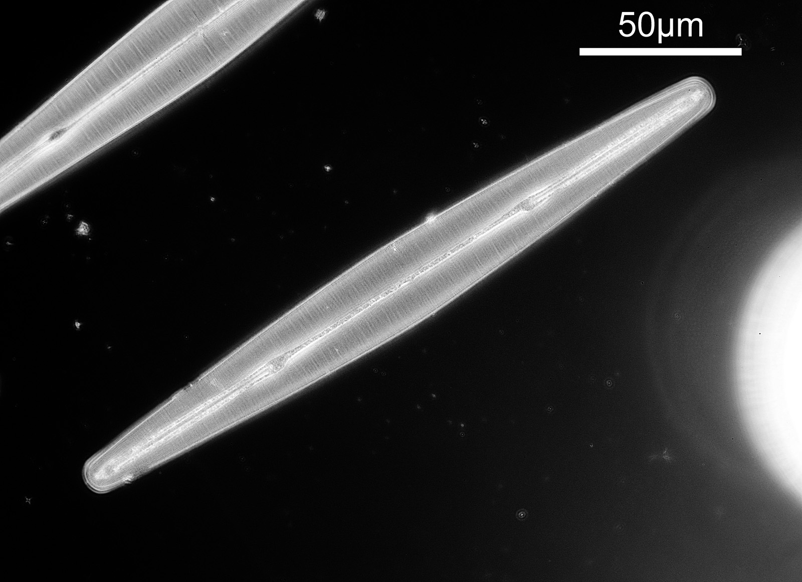
Enough chat, what do the images look like. Note, I’ve reduced the images resolution for sharing here. First with the Watson Holoscopic condenser.
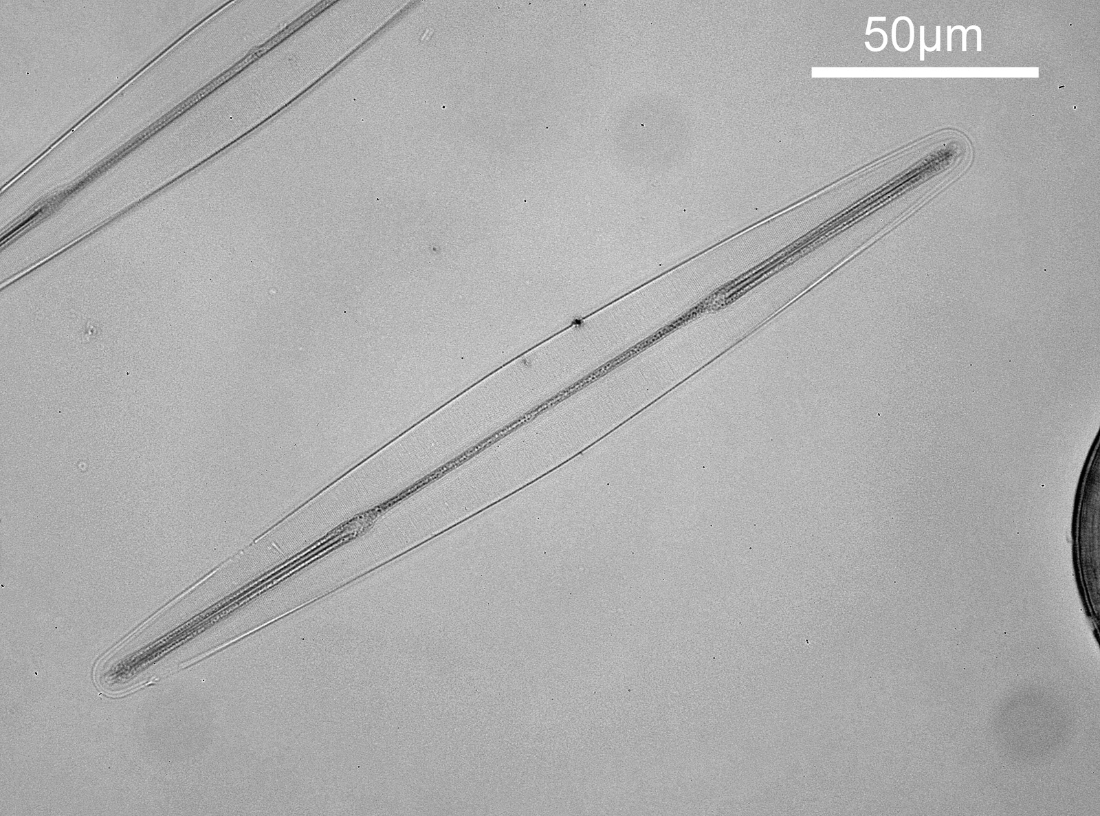
Next with an Olympus Aplanat Achromat condenser, in normal brightfield mode.
And finally, with the Olympus Aplanat Achromat condenser but in oblique mode.
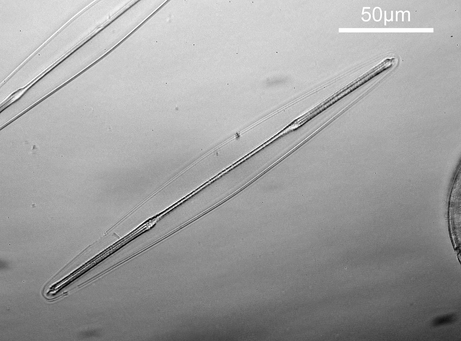
The Watson Holoscopic condenser gives dark ground (darkfield) appearance to the image, but for this to happen I had to stop down the iris on the 40x Olympus objective slightly – fully open it didn’t produce a dark ground image as the objective NA was too high. This is why it was important to use an objective with an adjustable iris for this experiment. It was probably just below NA 1.0. The iris on the objective was then left the same for the images with the Olympus Aplanat Achromat condenser. The low contrast of the normal bright field image can be seen, as well as the improvement in 3D appearance with the oblique condenser. In these full size images though the Watson Holoscopic condenser looks to show the features of the diatom quite well.
Crops of the 3 images show a bit more of the detail they can reveal.
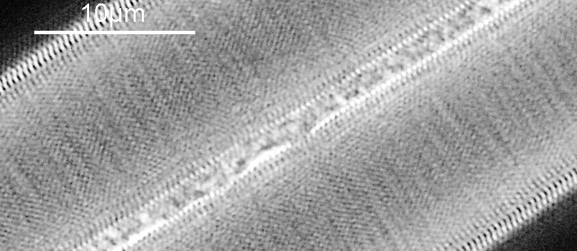
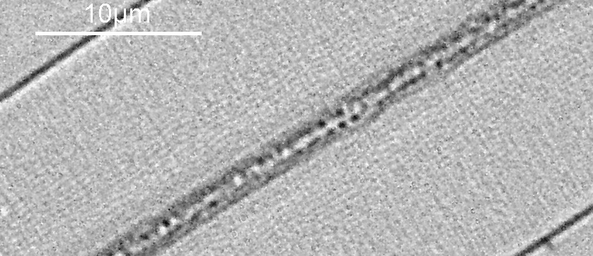

The benefits of oblique illumination over normal brightfield for the diatoms can clearly be seen in these cropped images. The Watson Holoscopic condenser also seems to be highlighting some of these features, especially at the edges of the diatom and along the central ridge.
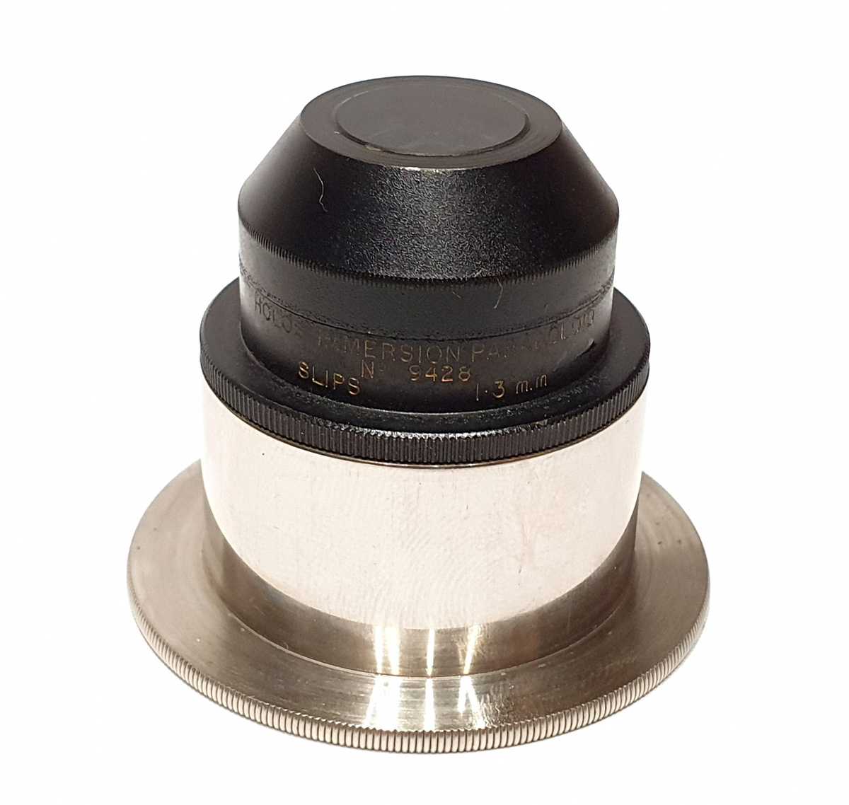
What is the Watson Holoscopic condenser? Here’s a picture of it.
And an excerpt from a 1930’s Watson catalogue which provides some information about it.
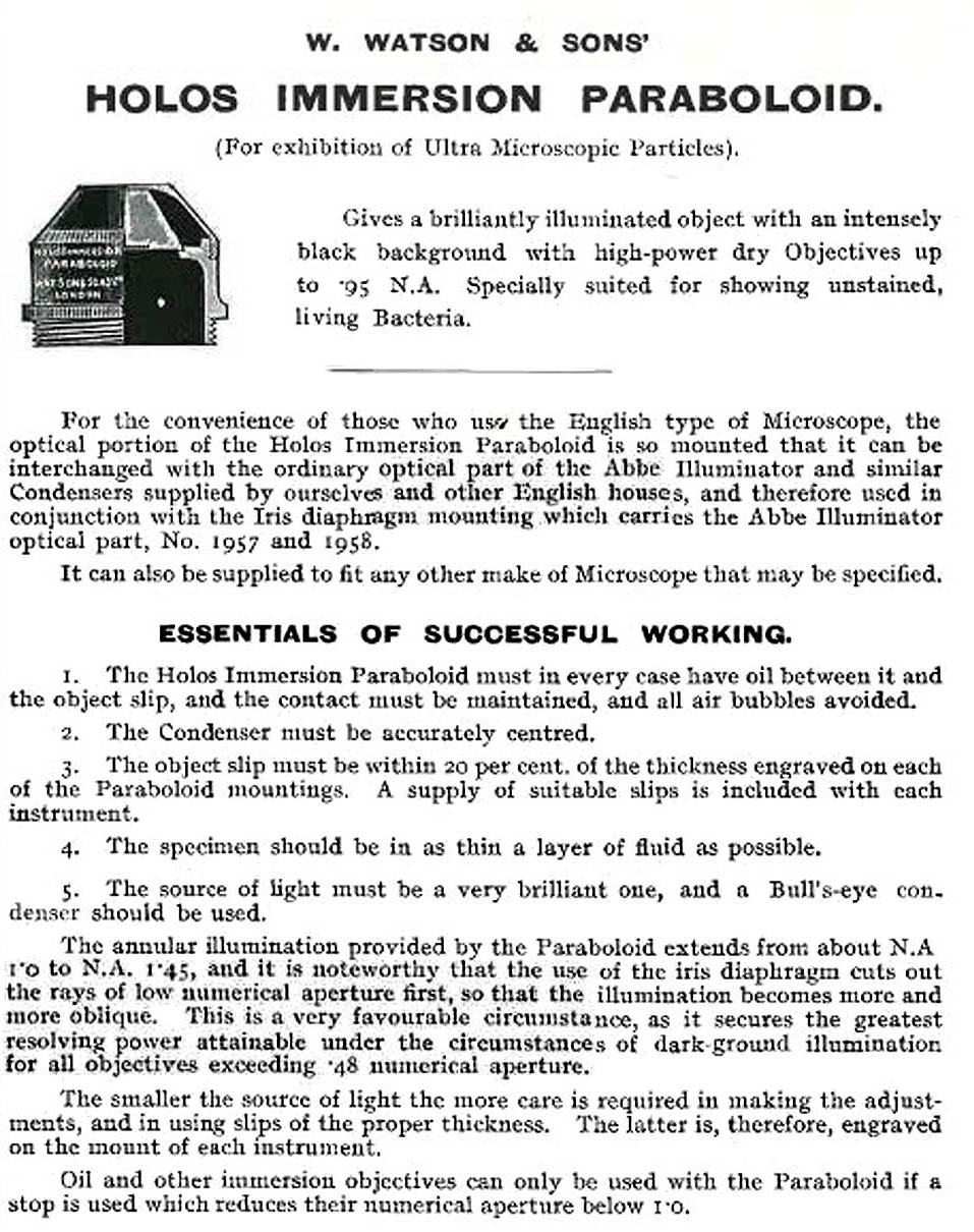
This shows why an adjustable aperture on the objective is useful, as with oil immersion objectives is requires an aperture of less than NA 1.0. It should be noted that the condensers were designed to be used with slides of specific thickness (this one being 1.3mm) so the slide used here was perhaps not ideal for this specific condenser.
The objective was quite an unusual one – a 40x Olympus UV FL PL NA 1.3 silicone immersion one with an adjustable iris.
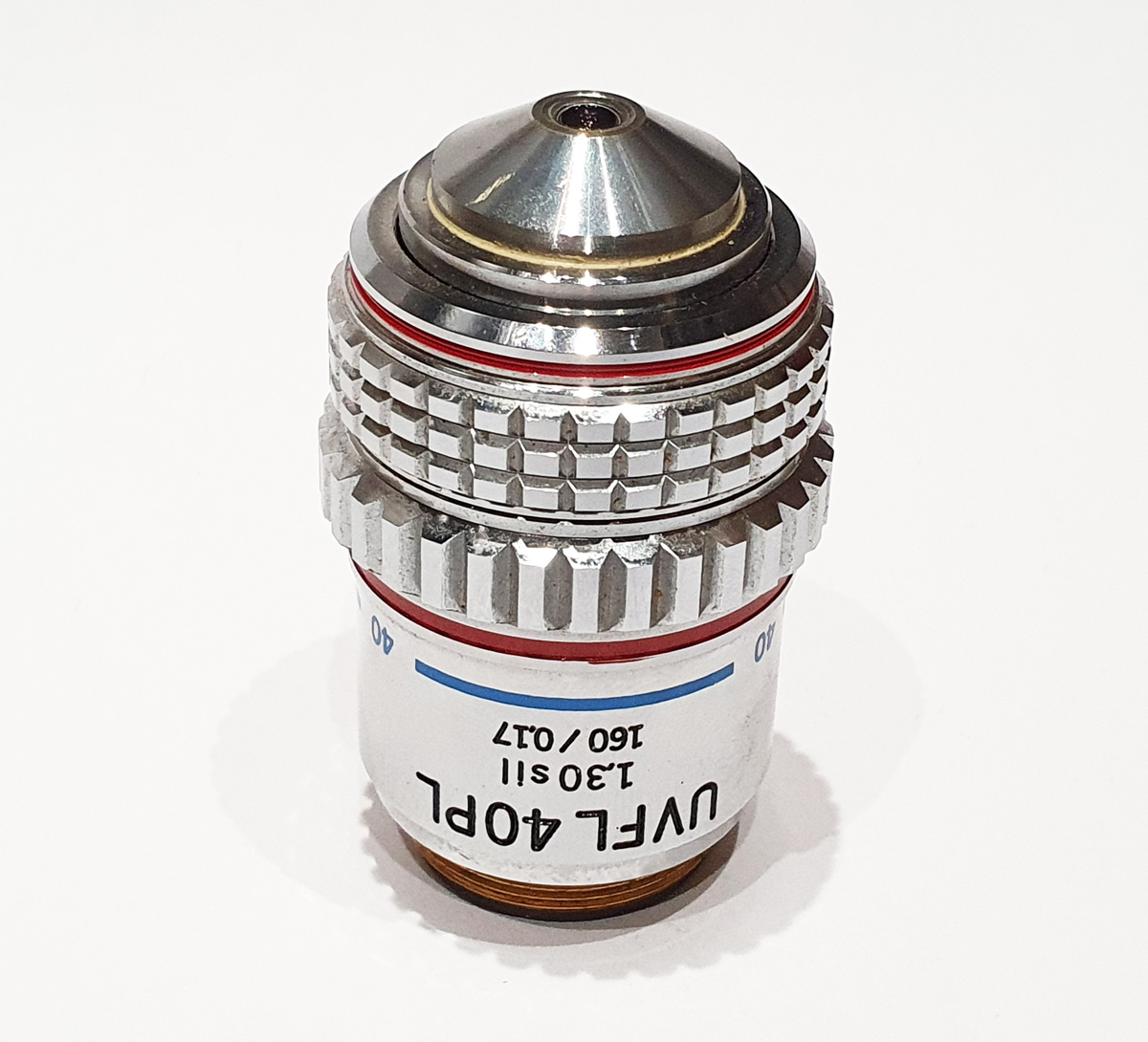
This is designed for UV fluorescence work, (‘UVFL’ on the barrel) so has quite good transmission in the upper UV region. It is also a phase contrast objective (the ‘PL’ on the barrel). It was meant to be used with silicone oil, although I just used normal immersion oil. I should have tried glycerine and water mix, as I have since read that that would be a better match to the silicone, but ‘every day is a school day’ as they say. Although not labelled as such it also has an adjustable iris to reduce its NA. This can be seen by looking through the objective when the knurled iris ring is rotated. This feature makes with useful for dark ground imaging, where tuning the objective NA to the condenser can optimize the image.
Finally a couple of shots of the slide itself. This was picked up on ebay for few pounds (GBP).

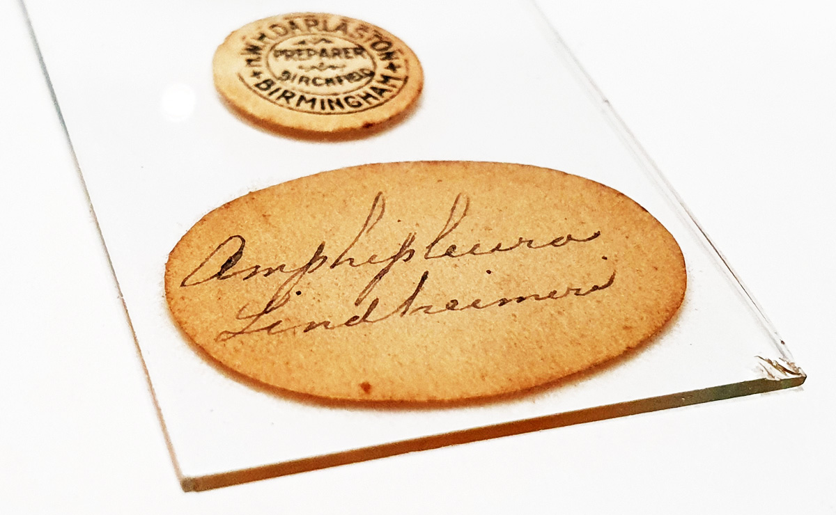
The original makes label says W.H. Darlaston of Birmingham, who is a well known slide maker. But is also labelled with T. Frankish, B.Sc, who I am guessing was the owner of the slide.
The Holoscopic condenser is a relatively simple construction, and I am wondering if it possible to make one of these using quartz or fused silica. If so it would be possible to use it deep into the UV which should allow for some very high resolution imaging. But that is for another day. As always thanks for reading, and if you’d like to know more about this or any other aspect of my work, I can be reached here.