
With my research interests in skin it was only a matter of time before I tried microscopy on a sample of it. Normally with skin sections they are stained to help bring out different parts of them. I wanted to try and see what an unstained sample looked like (the plan eventually is to look at it with UV, but that is a story for another day). Rather than using a new slide, I thought an antique one might be better and be unstained. I found an Edmund Wheeler slide of skin on ebay, and after a brief wait for the postal service to do its thing it arrived for imaging. Here’s the slide.
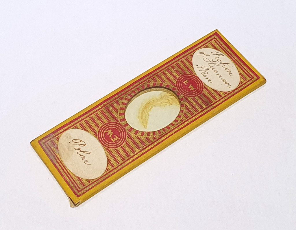
Edmund Wheeler (1808-1884) is a well known preparer of microscope slides. Indeed his work is often counterfeited, although I am reasonably confident that this one is not a fake and is probably around 150 years old. As you can see in the middle of the slide is a large section of skin which looks slightly dark against the white background. How does it look under the microscope? Firstly with normal bright field imaging. This was with a 10x Olympus UVFL objective and a tungsten light source and a scale bar is included.
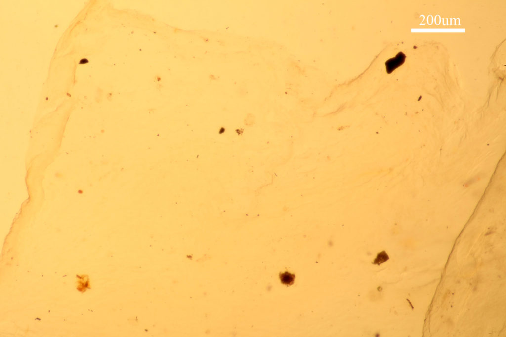
It’s pretty obvious with bright field imaging and an unstained skin sample, it is difficult to see anything. You can just about make out the edge of the skin, and that is about it for structures.
Switch to phase contrast and everything becomes clearer. This is the same region, but now with a 10x phase contrast objective and lighting.
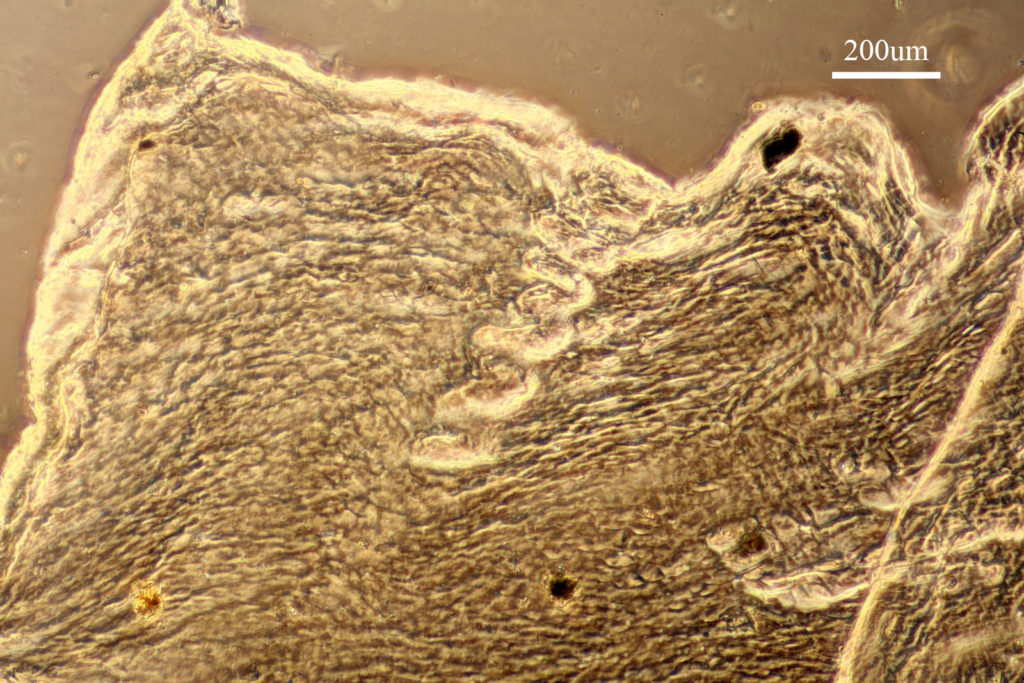
Using phase contrast a structure appears in the middle of the image, with the corkscrew cross section of a sweat gland. In fact looking around the sample there were a few of these features moving down into the skin from the surface. There are also different layers of the skin which are clearly defined, so I’ll be exploring that more in the future.
Microscopy is an amazing tool for exploring the natural world, but as with all imaging techniques, choosing the right setup is key to actually being able to see what you are interested in. Here, phase contrast imaging has revealed features of an unstained skin sample not possible to view using standard bright field imaging. If you’d like to know more about this or my other research you can contact me here. Thanks for reading.