
One of the really fascinating aspects of my microscope refurbishment project has been the ability to look at things which are impossible to see with the naked eye. This is of course because of the magnification it can offer, but also the ability to play with the lighting to emphasize different aspects of the subject. The ability to look the natural world with a microscope has fascinated scientists and artists since microscopy began. The Victorians had a particular fascination with something called Diatoms (the silica shells formed by microscopic algae), making amazingly beautiful patterned slides comprising tens or hundreds of them, and there’s a great article on that here. These old slides are extremely sought after, and command huge prices, however less ornate vintage slides with diatoms on them regularly appear on ebay and other auctions sites, and I thought it would be great to get one and see what it looked like using my microscope build – Project Beater.
The slide I got was made by Clarke and Page, who worked in London between 1904-1923 (there’s a bit on history on them here), and it only had a few diatoms on it. On the plus side it cost me less than 10GBP. So, the important stuff, what does it look like? All images below were taken using a camera phone through the eyepiece, and have not been focus stacked. Firstly imaged using 10x objective, and 10x eyepieces for 100x overall magnification with standard brightfield imaging.
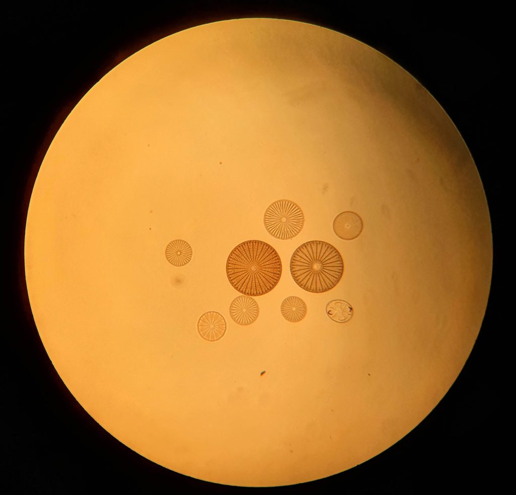
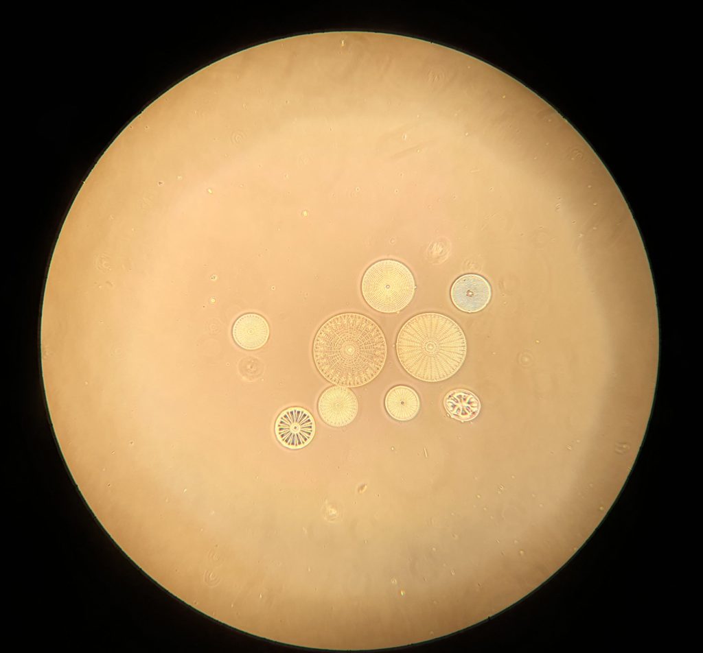
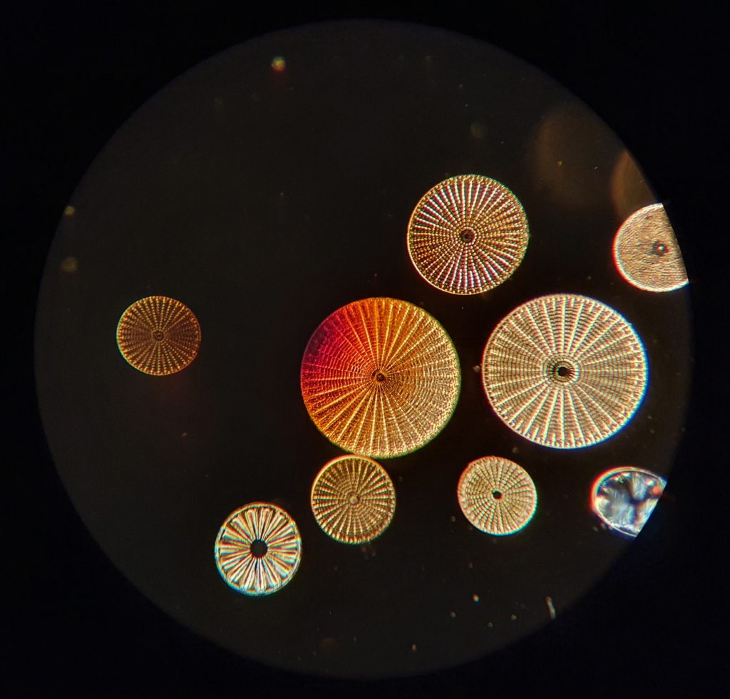
Even with brightfield imaging at this magnification the structures present inside the diatoms is starting to become apparent. Where diatoms come ‘alive’ though, is when you do phase contrast and darkfield imaging. Here they are under phase contrast and darkfield imaging at the same magnification.
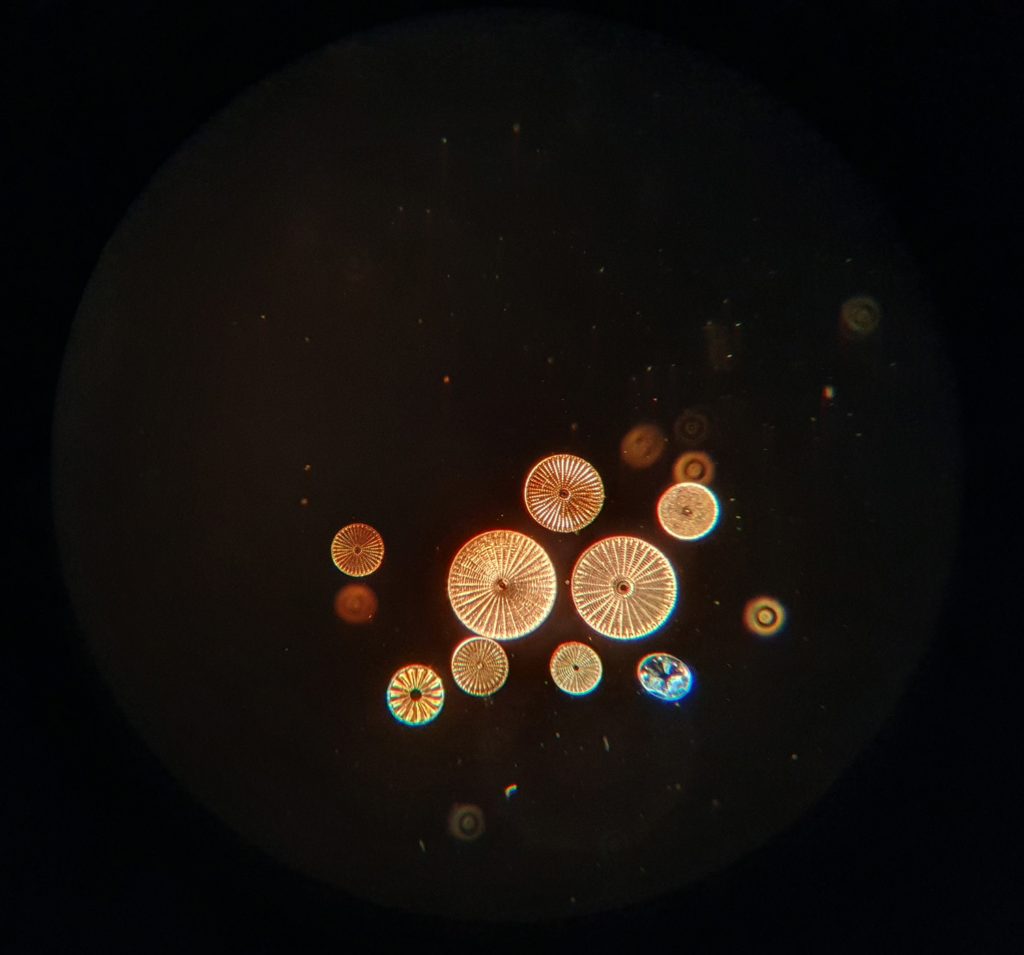
As you can see, they looks much more interesting when the lighting is changed. I also took a couple of images with a 20x objective, for 200x overall magnification – brightfield and darkfield.
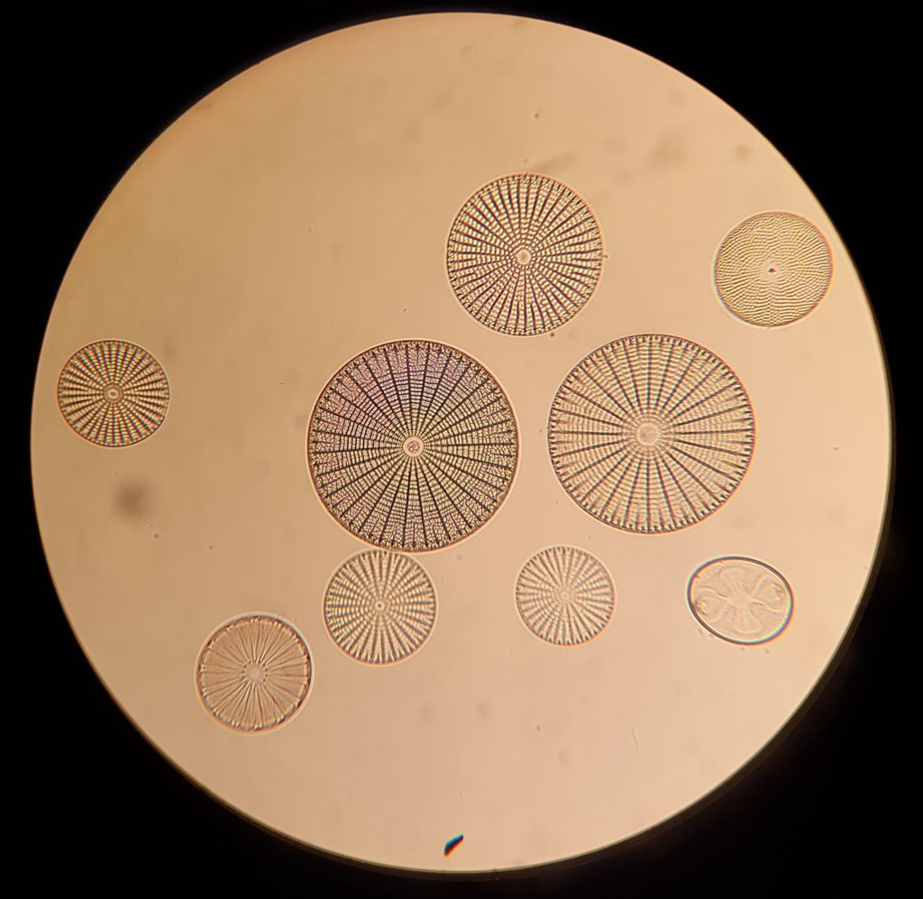
These images were just taken with a camera phone through the eyepiece, so the quality is obviously poor compared to focus stacked images, and as I learn more about getting good quality focus stacked photos, this slide will be a great subject to have a play with.
Here’s the slide itself.
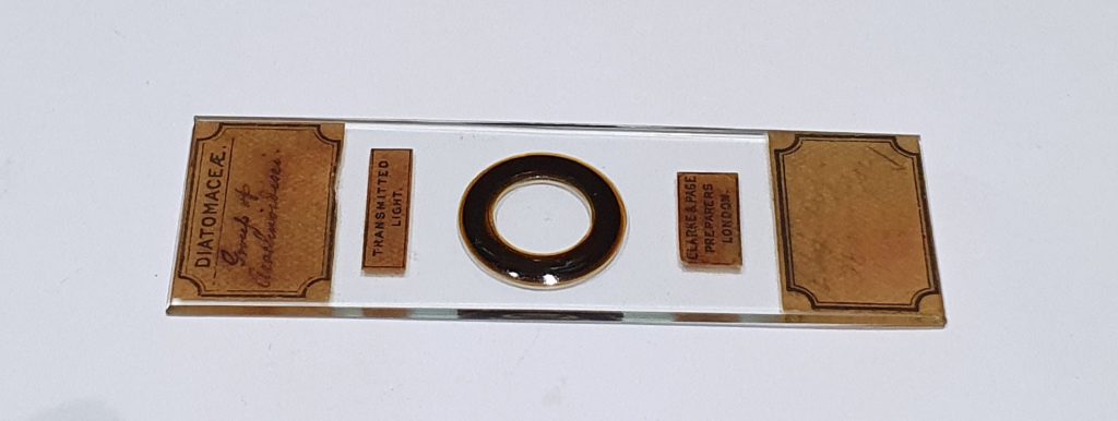
The diatoms themselves are just off the center of the black ring in the middle of the slide. Depending on how good you monitor and lighting are, you may just be able to make out a tiny dot just off center within the ring – that is the cluster of diatoms.
Using a microscope can literally open up a new world to anyone interested in science and nature, and for not much money you can walk in the footsteps of the early pioneers, seeing what they saw. Rebuilding a microscope has offered me fascinating insight into this world, and is something I will continue to research both for work and pleasure in the future. If you want to know more about this or any other areas of my work, you can reach me here.