
On the 12th August 2023 I attended Microscopium which is an annual event held by the Quekett Microscopical Club where members and visitors meet for the buying and selling of surplus microscopy related items. There were some fascinating items for sale, including some absolutely amazing microscopes, and I came away with a couple of boxes of microscope slides of diatoms (and other things) which I am now gradually working through. Today I’d like to share some of the initial images of the slides.
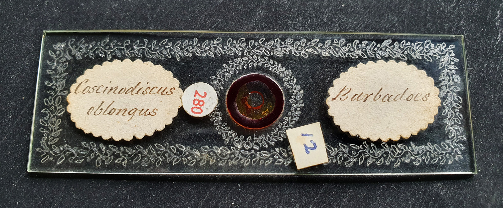
First is a slide by Frederick Marshall of three Coscinodiscus oblongus diatoms. This slide is beautifully decorated with an engraved pattern all over its surface.
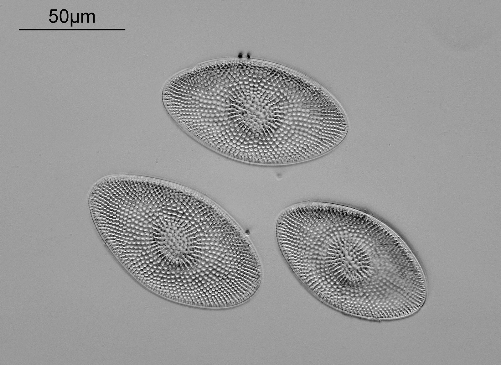
The diatoms from the slide are shown below.
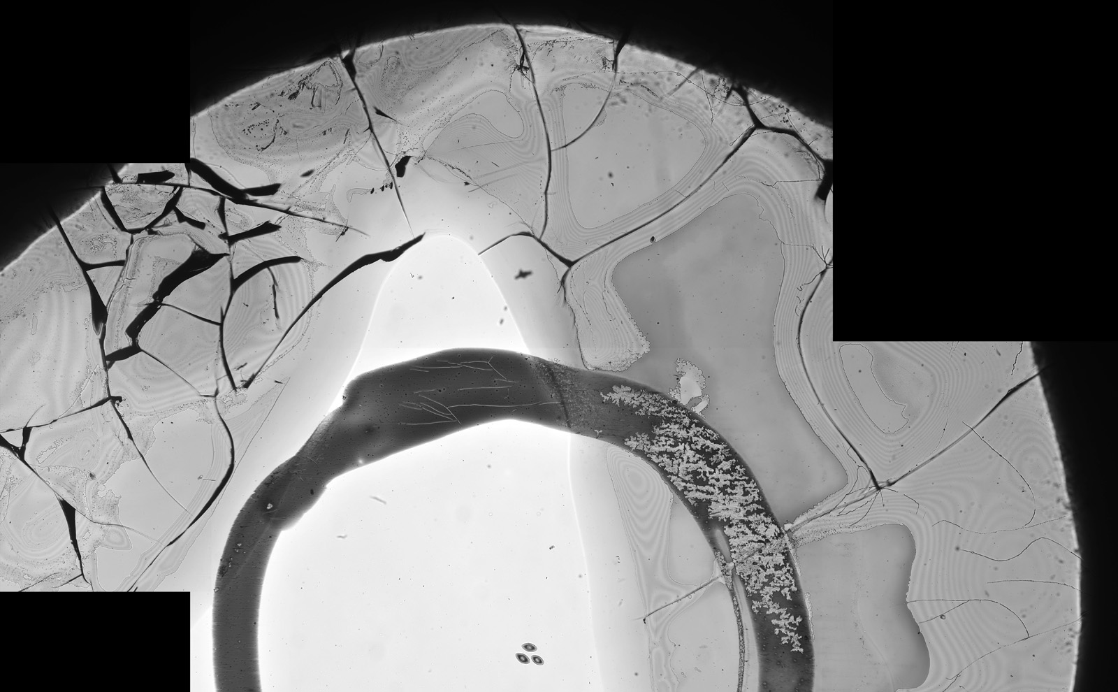
Unfortunately at some point in its life, the coverslip has been damaged, as shown in the composite image below.
It turns out that these engraved Frederick Marshall slides are quite rare and desirable, and I got lucky with this one as it was in with a box of 100 diatom slides. For more about Frederick Marshall see here.
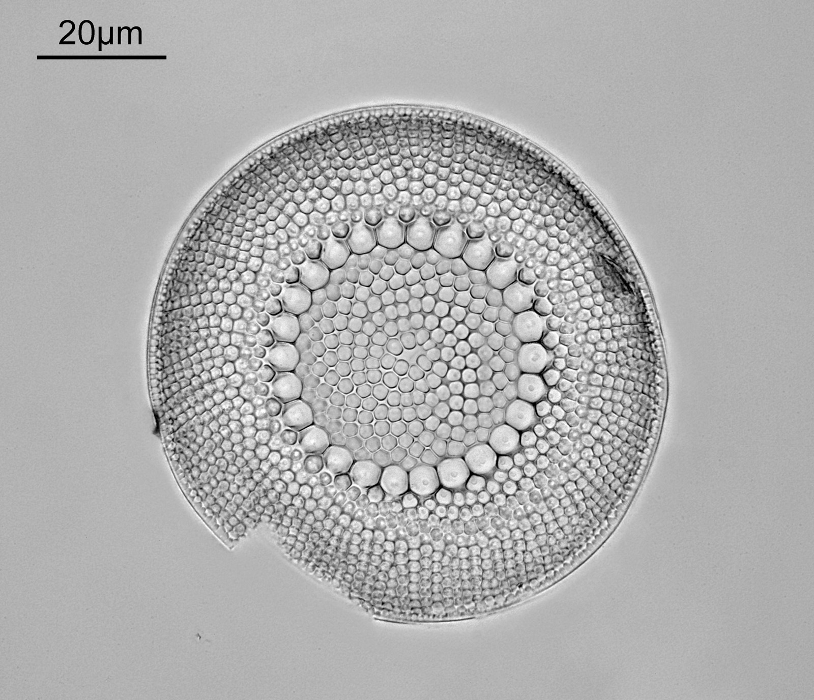
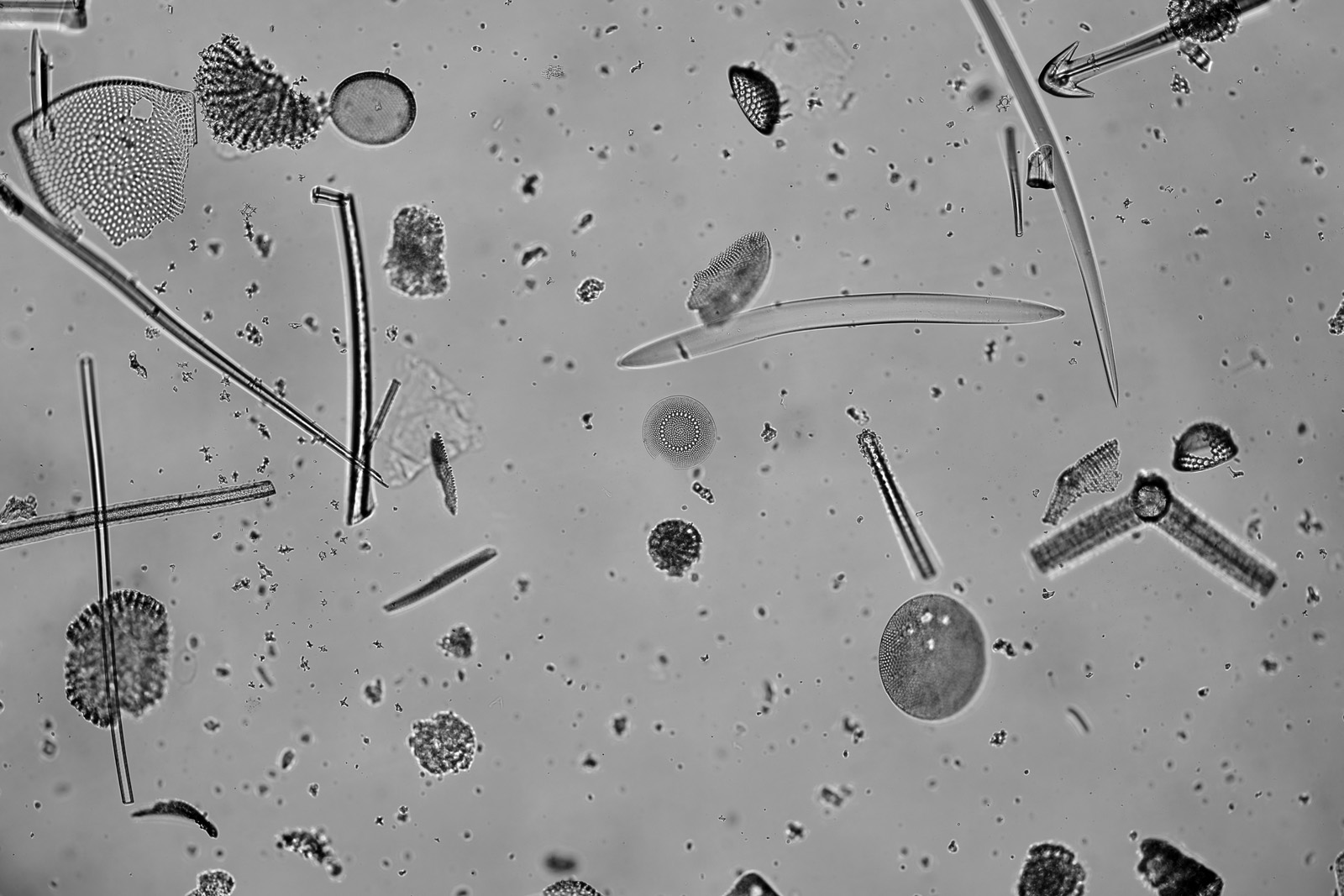
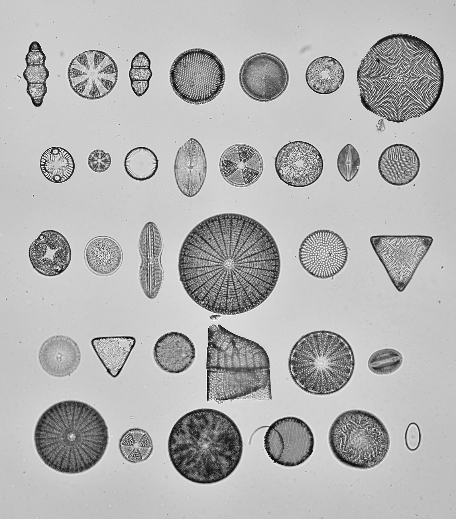
Next is a Brightwellia coronata diatom from a strew slide from Jackson’s Paddock, Oamaru, New Zealand.
This is a strew slide with a lot of material present, as can be seen from a wider field of view below (B. coronata in the middle of the image).
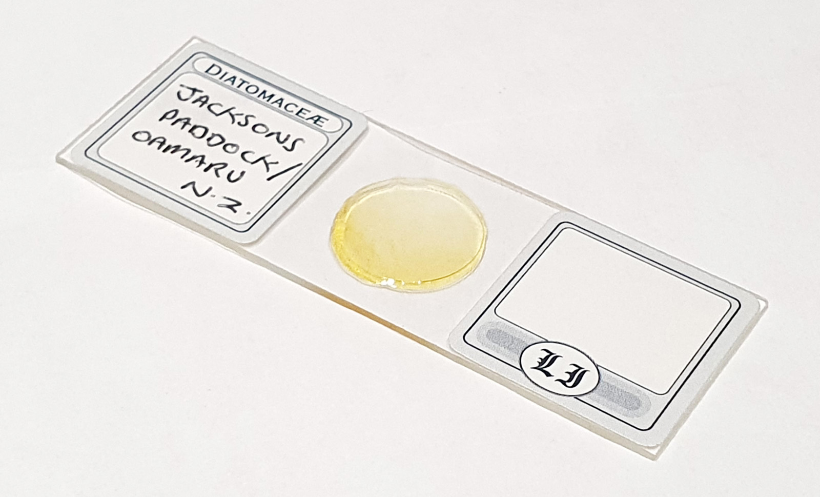
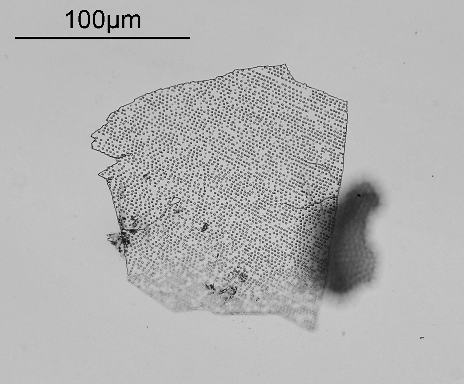
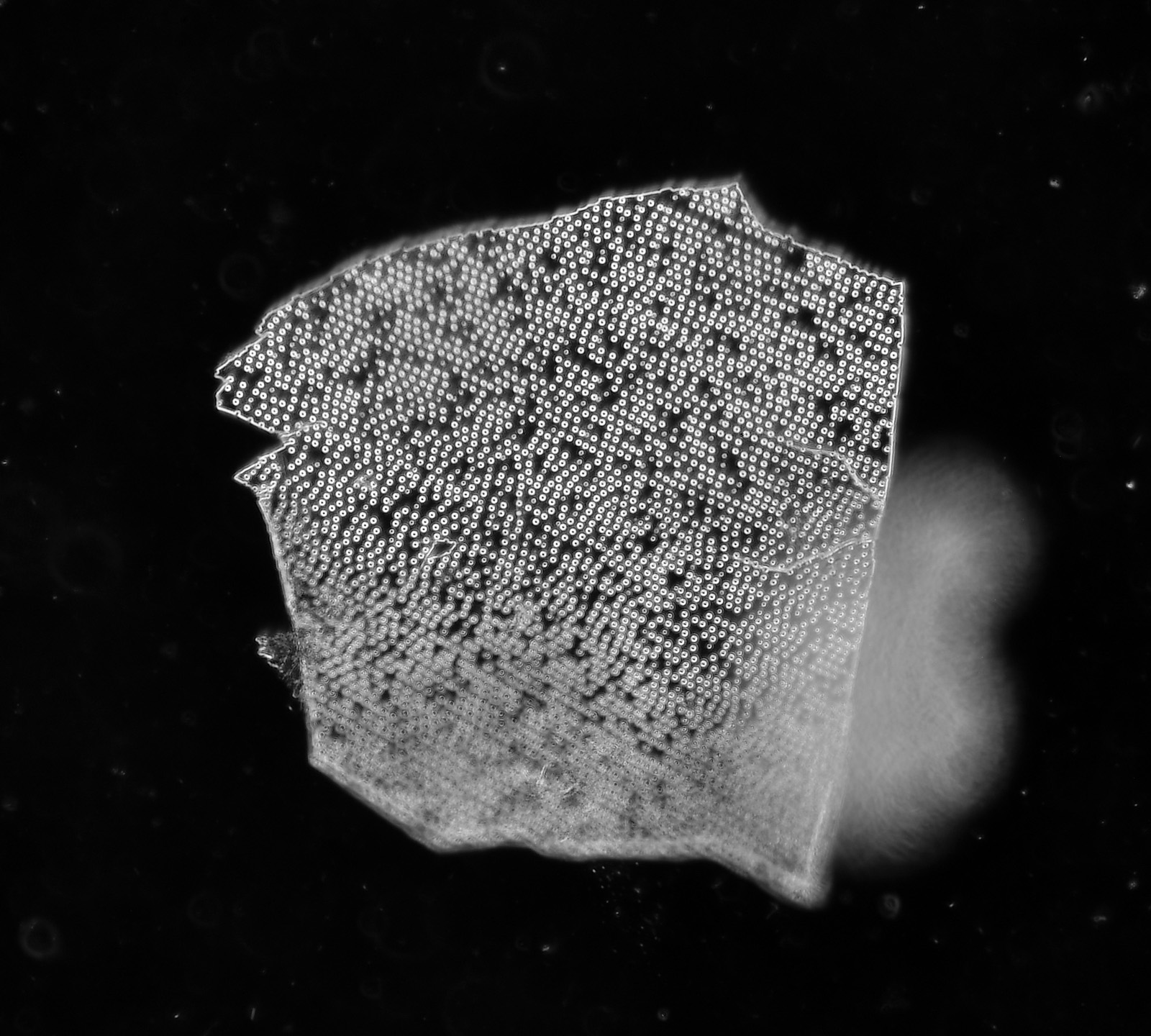
I’ve yet to find an intact one of these on a slide, and this is about the most complete one I have come across so far. Apparently the ‘LI’ on the right hand side stands for ‘Little Imp’. An image of the slide is given below.
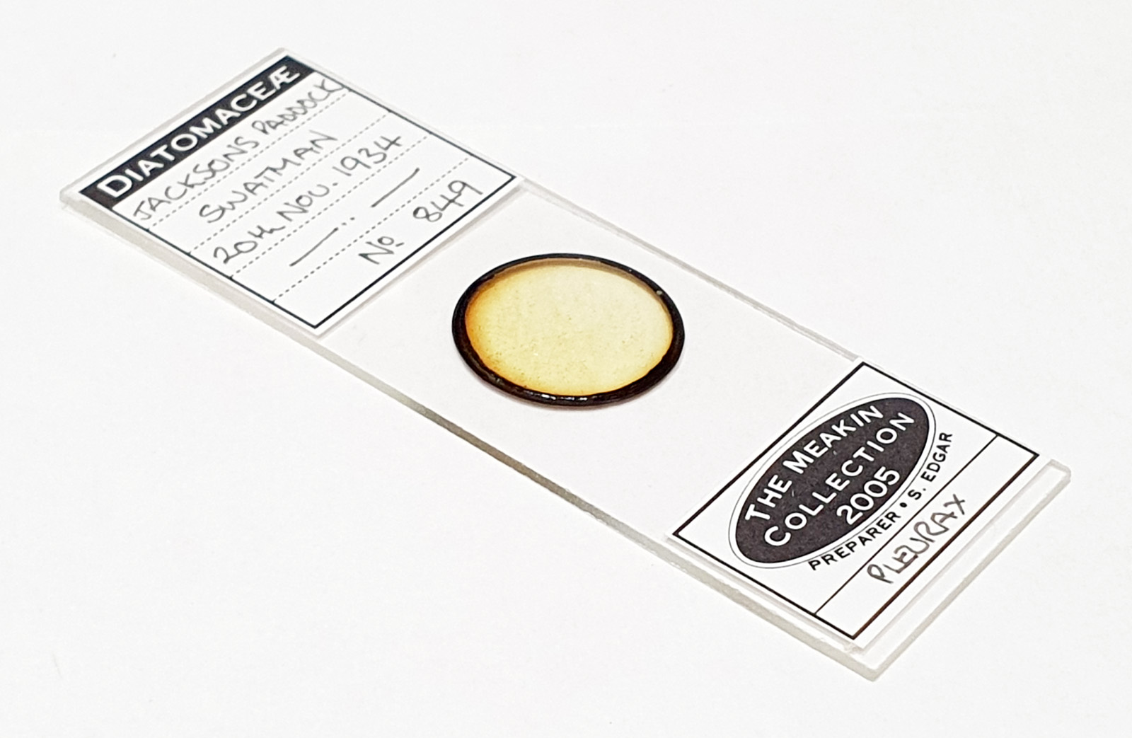
The next one is an interesting curiosity, and also from a Jackson’s Paddock strew slide, although this one was made by Steve Edgar as one of his Meakin Collection slides.
I’m not entirely sure what this is, and I initially thought this was something artificial and a contaminant, although having spoken with a diatom expert it could be part of a sponge spicule. It looks really cool in dark ground imaging.
It reminded me of a punched card and I went back and did a high resolution image using a 63x Leitz Pl Apo NA 1.4 objective and a stack in Zerene.
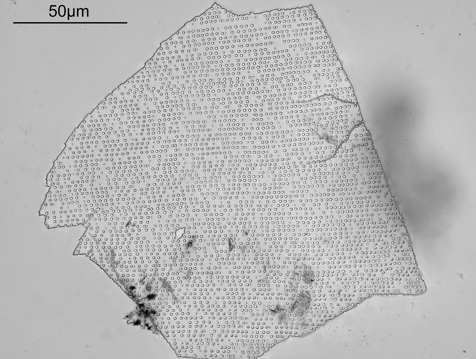
It still amazes me as to how artificial this looks. Very curious and I shall look out for similar examples in other slides. Speaking of which here’s the slide it came from.
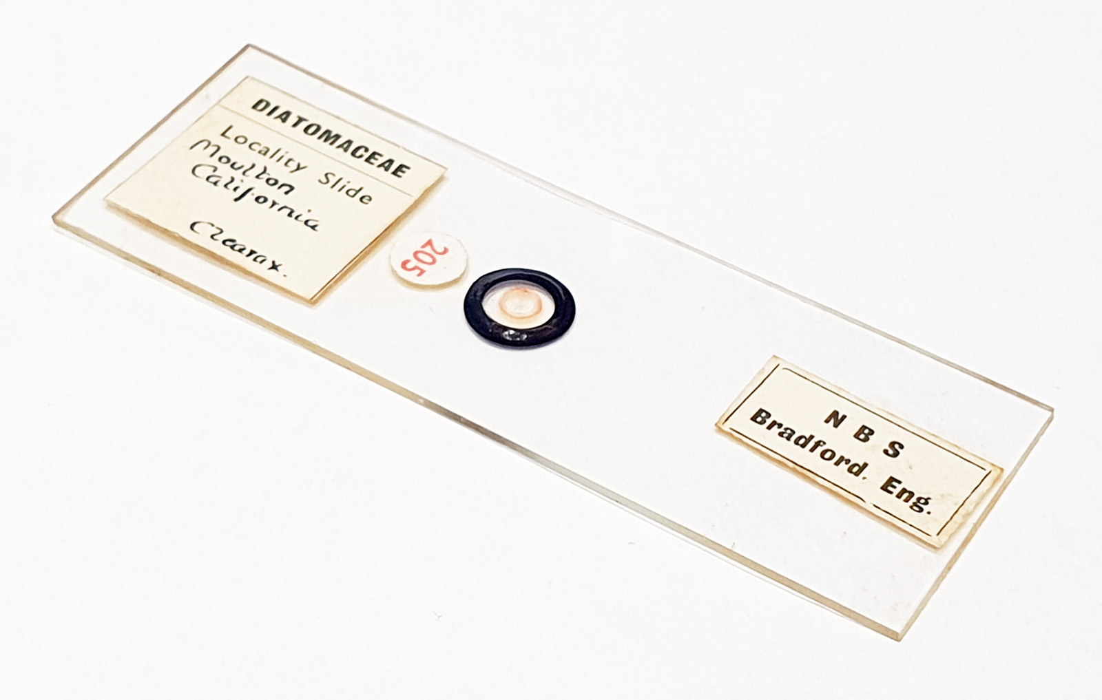
Next is a locality slide from Moulton, California made by Northern Biological Supplies (NBS).
And the slide itself.
The image I took of this was not stacked, and I’ll return to it when I have more time to do a better one.
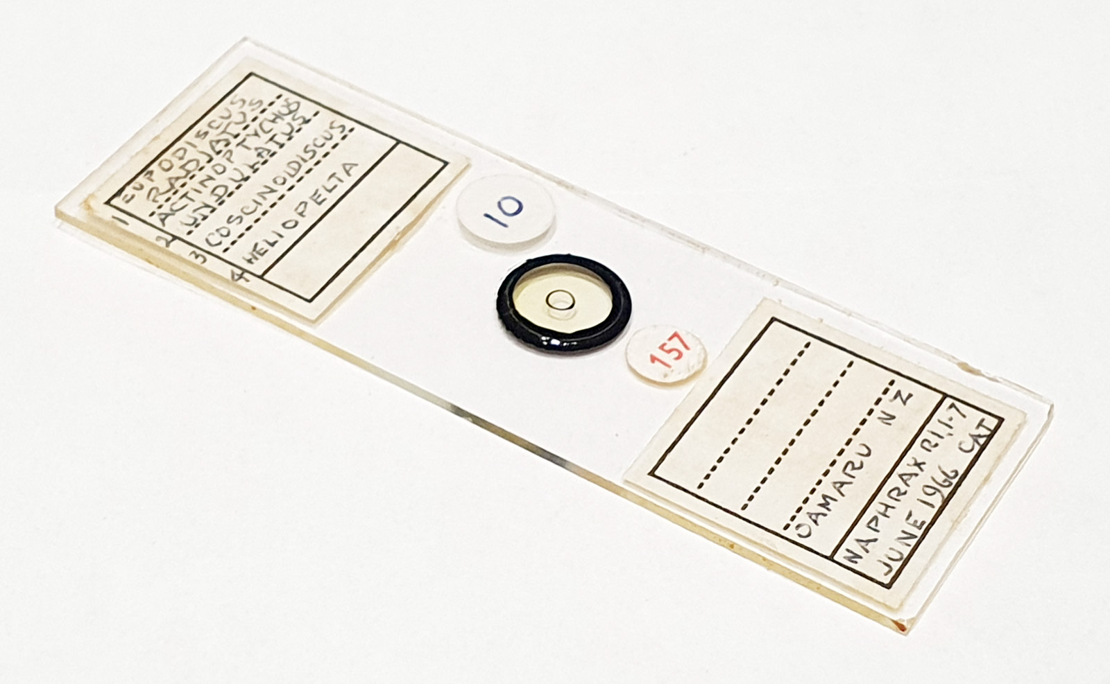
Finally for now, back to Oamaru, and a 4 form slide (unknown maker).
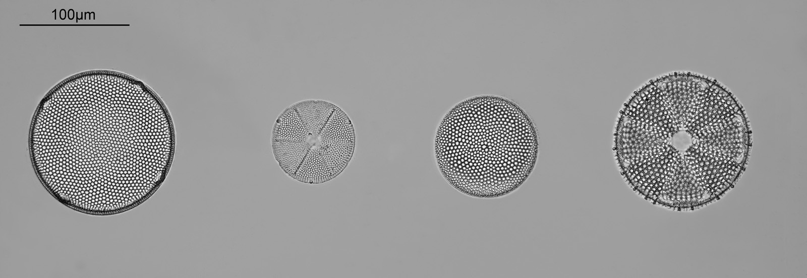
Here’s the slide (if you know the maker drop me a message please).
These were some of the nicest slides I have found so far from the Microscopium event. Most of the ones I got were strews, and they aren’t all like these, but even so I’m more than happy with what I managed to find there. I also came away with a box of slides of different Bacteria which it will be interesting to look through as they are completely different to my usual subjects.
The Microscopium event was great and although you don’t need to be a Quekett member to attend I can well recommend membership to the club. There is an incredible depth of knowledge in the club and the USB copy of the old Quekett Journals is well worth getting. For a summary of this years Microscopium event see here. One thing I would say about Microscopium it is not for people to go along and pick up bargains to then subsequently sell, this is for those passionate about microscopy.
As always, thanks for reading and I hope you enjoyed the images. If you’d like to know more about my work I can be reached here.