
A quick update for this post, looking at some work I’ve been doing with UV microscopy recently. I’ve been imaging a strew slide by Neville Bradpiece made in 1998 which has a wide range of diatoms on it from Toome Bridge, Northern Ireland. Today’s post looks at some Pinnularia diatoms, and imaging of very small features – poroids – in their structure using 365nm UV light.
First an unstacked image of a Pinnularia from the slide, taken using my modified Olympus BHB microscope and a 365nm LED light source (image is shown at original pixel resolution).
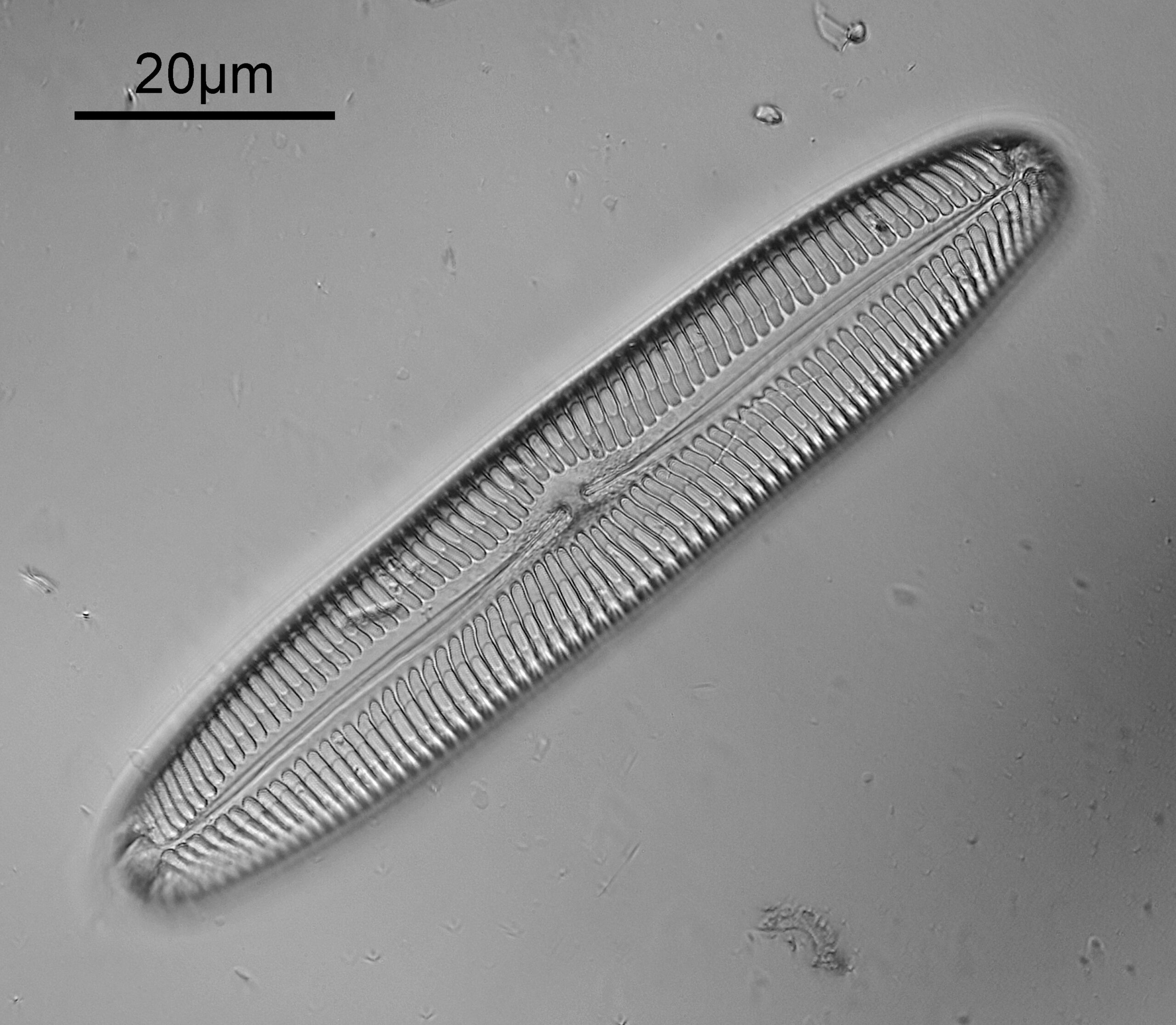
Going in close to the top right of the image starts to show a hint of of the poroids as dots in the structure.
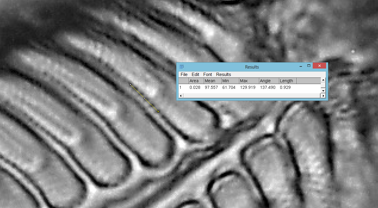
Using ImageJ, I measured 929nm between 6 of them, giving a spacing of 186nm centre to centre. Poroid imaging in Pinnularia is seen as a huge challenge, given how tiny these features are, and it is normally recommended to use circular oblique lighting and cross polarization. This image was done using bright field with slightly (linear) oblique lighting using a Lomo OI-14 Aplanat condenser, and no polarization. The objective was a 63x Leitz Pl Apo NA 1.40, with oil immersion. The condenser also used oil immersion. The benefit here comes from using 365nm UV light, with the shorter wavelength offering improved resolution.
Even in my optimistic frame of mind would accept that the image above barely has enough resolution to see the poroids, however I did image are few other examples of Pinnularia on the slide, one example of which is shown below.
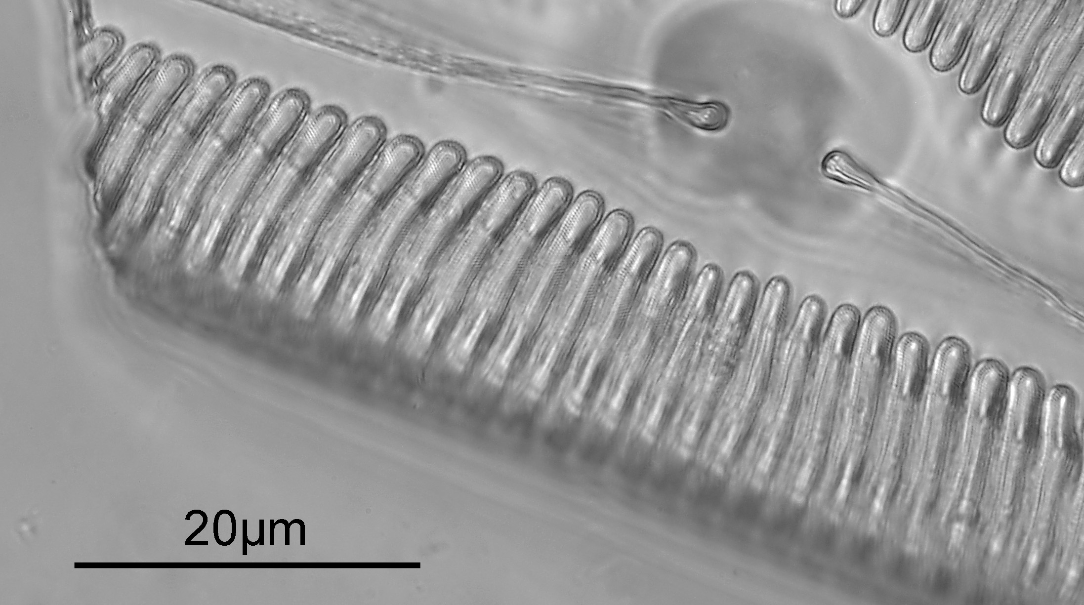
Going in close and doing some measurements with ImageJ gave the following.
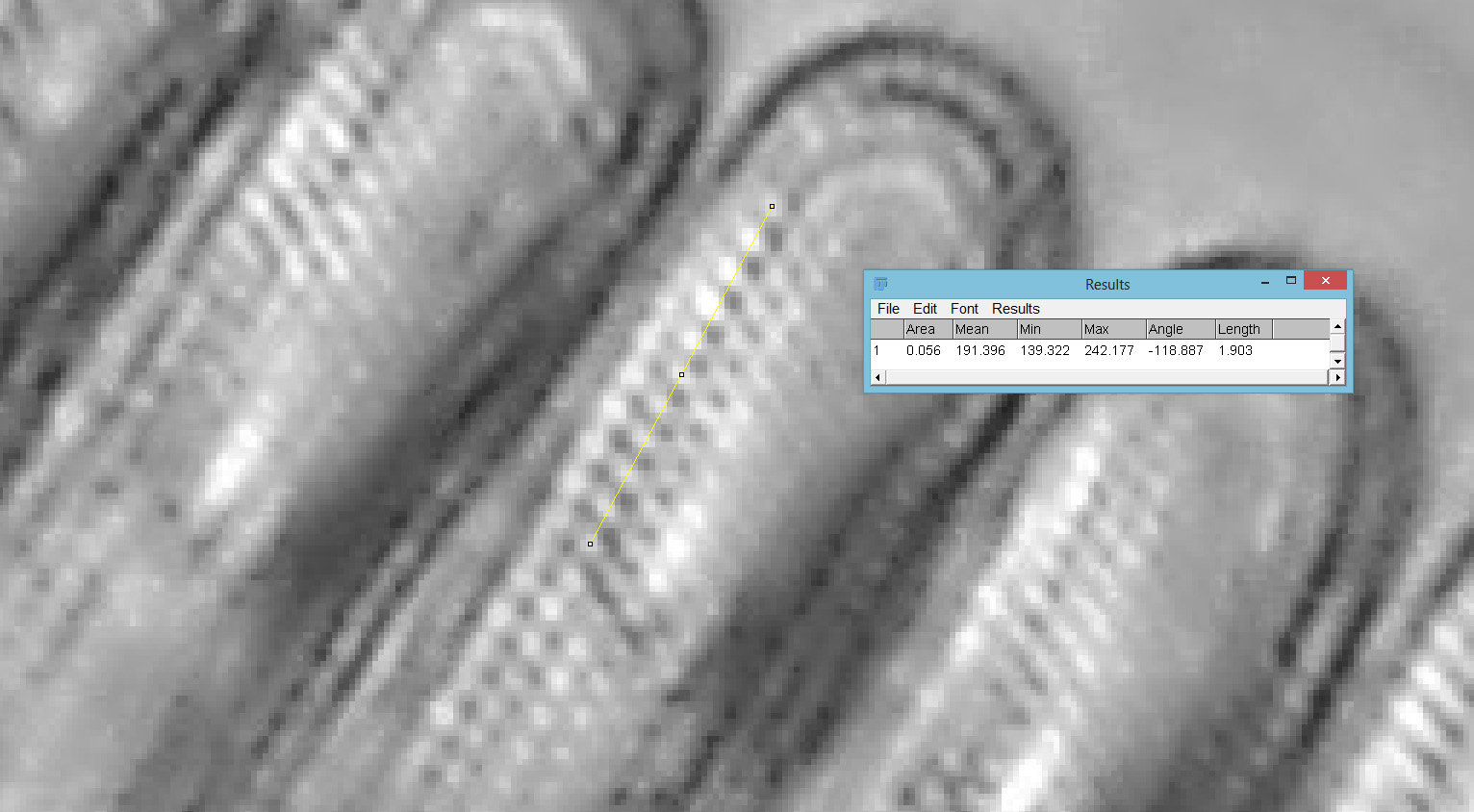
With this one, the spacing worked out as 190nm centre to centre, and they are a bit clearer here. Other examples gave poroid distances of 235nm and 251nm so there was a bit of variation between samples.
As always, an image of the slide made in 1998.
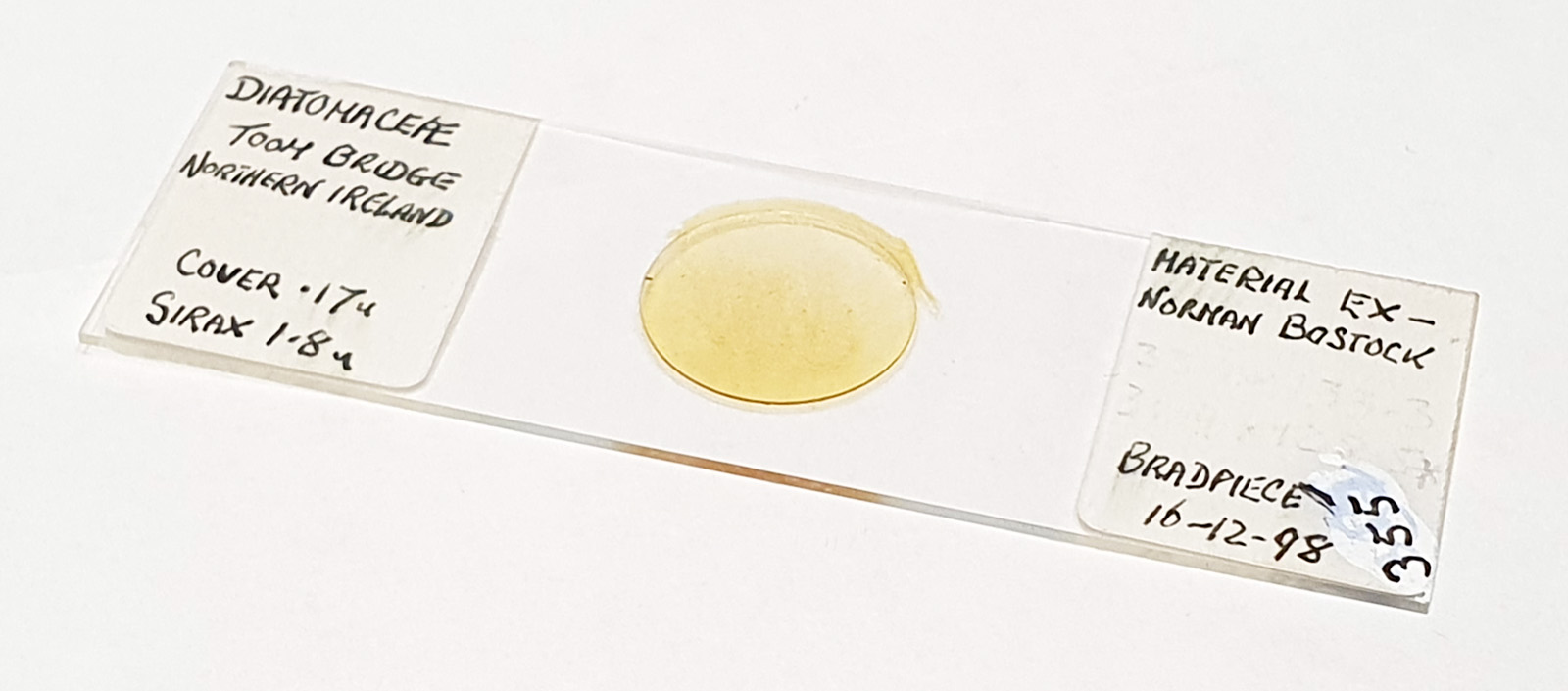
The slide uses Sirax as the mountant and quotes a refractive index of 1.8(!). Sirax refractive index is quoted between 1.65 and 1.81 depending on sources, and I’ll be getting more into this when I get round to writing a full article on this slide. High refractive index is great for imaging diatoms due to the improved contrast it offers. There are lots and lots of different diatoms on this slide, and I think it warrants a proper paper showing more images from it. Another article for the list of ones to write…..
As always, thanks for reading, and if you’d like to know more about my work I can be reached here.