
I have an unusual (and rather rare) diatom slide to share with you today – an example of Hydrosilicon mitra, prepared by S.H. Meakin. This was made in 1933, and is mounted in Hyrax (which will become important later). The images shared were taken on my modified Olympus BHB microscope, with a monochrome converted Nikon d800 camera. The images have been reduced in resolution for sharing.
The first is a simple bright field image. This is a stack of 7 images and was done using 450nm filtered LED light. The objective was a 40x Olympus Dplan Apo UV (NA 0.85). Condenser was an Olympus Aplanat Achromat.
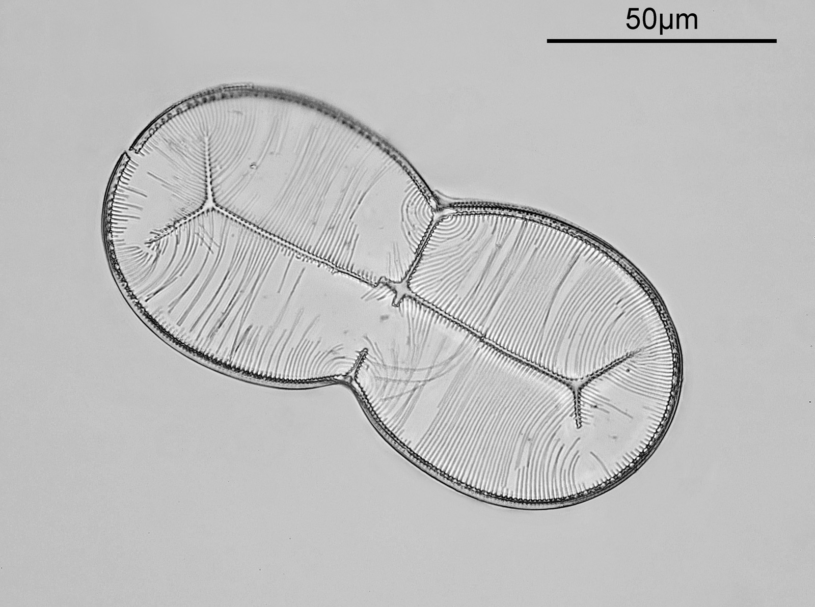
The sample is not flat, and dips towards one side. This means that stacking was necessary with the 40x objective (in fact I missed the focus on the top left of the diatom – ” ‘B-‘ must do better next time”). As can be seen there is some damage to the fine structure of the diatom, but it is still a very pretty specimen.
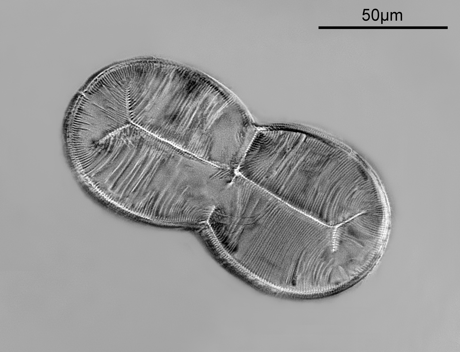
I wanted to see what this looked like with dark ground illumination, so I got out my Watson Holoscopic condenser (some info about that here) and used glycerine as the immersion fluid to attach it to the underside of the slide. The objective was a 20x Olympus Splan NA 0.46, and again 450nm light was used. No stacking this time, just a single image.

I love the dark ground effect, but being a single image part of the diatom is out of focus due to it not being flat. I’ve not got my head around stacking dark ground images yet.
As a final image, I wanted to try something different. As mentioned above the mount is Hyrax, and this has pretty good UV transmission at 365nm. I wanted to try imaging with 365nm light (to potentially help improve resolution), and also use a different method of illumination – circular oblique light (COL), also known as annular illumination. To do this I used a 40x Olympus UVFL PL NA 1.3 silicone immersion objective in combination with the Watson Holoscopic condenser. This objective has an adjustable iris – wide open and the NA is bigger than that of the condenser and the image is bright field. Iris closed all the way down and the image becomes dark ground as the objective NA is now smaller than the condenser. In the middle ground COL can be produced, when the NA of the objective is close to that of the condenser. This gives a nice 3D effect to the image. Both the condenser and the objective were used with glycerine as the immersion fluid. The image below was a stack of 13 images, using 365nm LED light, and with a COL setup.
COL illumination gives a more 3D effect to some of the features. But, and this is a big ‘but’, the resulting image was very noisy, and was a bit of a nightmare to stack. Not sure why it was so noisy, as I used ISO200 on the camera. I really like the end result though and will definitely try this technique again.
As always, here’s the slide itself as well.

Not too bad for a slide made 90 years ago. The slide was bought on ebay for about £30 and there is a single example of the diatom on it. As always, thanks for reading, and if you’d like to know more about my work, I can be reached here.