
Sometimes I see something for sale, and despite not knowing quite why, know that it’ll be interesting or useful to have. So it was the other day when I saw a microscope slide for sale. This was a diatom slide made in 1950 by a chap called Horace Dall, but what caught my eye was that the slide said that the diatoms were mounted in titanium dioxide (TiO2), and then it gave a refractive index measurement – ‘µ = 2.90’. Before I show you the slide though, I want to share an image taken of it, as it turns out it makes for some really spectacular photos.

I’ll speak more about the images from it in a minute. Back to the slide itself. Here’s a photo of the slide;
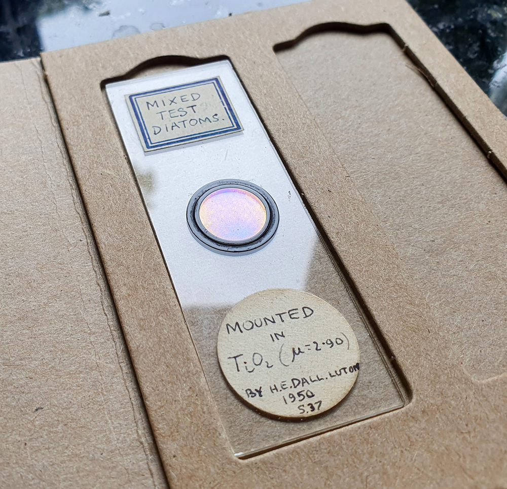
I’d not seen anything like this before, and guessed that it had been done to improve contrast of the diatoms for imaging (given the high refractive index of the TiO2). The sample under the coverslip had a sheen to it a bit like an oil film on water. After a bit of a bidding war I ended up with it and the imaging began. As a simple set of initial images I did some brightfield and darkfield ones with a 20x objective (Olympus Splan 20x) and a 63x objective (Zeiss Neofluar Pol 63x) as I could use both of these without needing to mess around with immersion fluids. Images were captured using a white LED light, and with a normal unmodified colour camera. The colours you see below have not been modified by me, these are as captured by the camera. Anyway, less chat, more images;
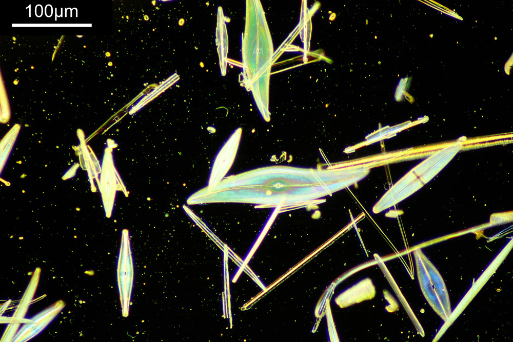
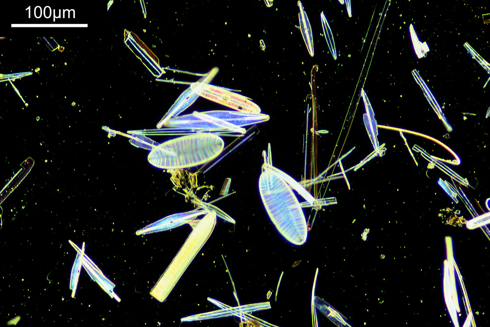
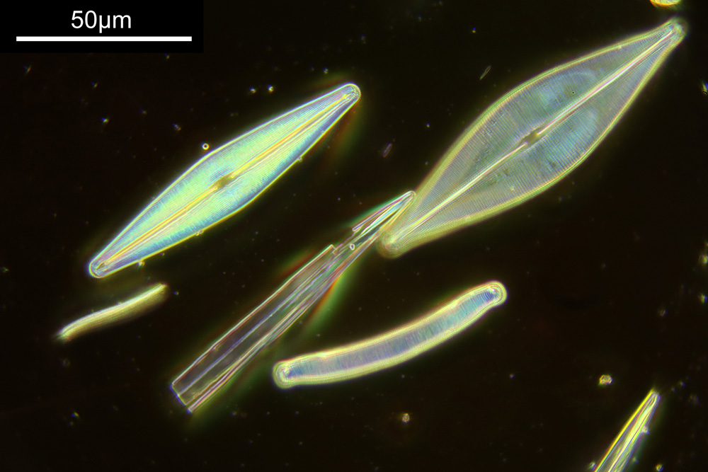
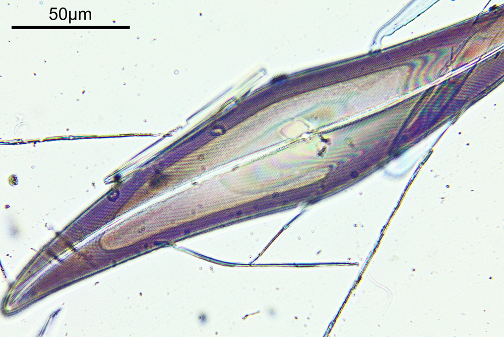
The darkfield images especially were very pretty, showing a range of colours from blue through to red for different diatoms. Even the brightfield image showed the range of colours. The effects reminded me a bit of looking at mica particles which are put in cosmetics, or an oil film on water, and I presume this is due to the thin layer of TiO2 deposited on the diatoms causing interference colours. This led me to wondering whether anything had been published on this process or these samples. I spend my days tracking down information on stuff, so set about looking for details on this, and, ……..nothing. So I approached the Facebook group of the Quekett Microscopical Club that I am a member of, and within an hour loads of useful information came back, including reference to a paper in the Quekett Journal from 1985 written by Horace Dall about making these types of slides. So, I need to learn more about searching for information….
Horace made these slides by exposing them to fuming titanium tetrachloride, which was then transformed into TiO2. Back when I was a student in Durham I won the Tioxide Scholarship for the year during my Chemistry degree. This came with a trip around the Tioxide plant on Teesside, where I was given horror stories about titanium tetrachloride and how dangerous it is. In Horace’s paper, he mentions “These experiments were carried out only when suitable winds would quickly evacuate the fumes.”. Legend. After this he went on to develop other approaches including the evaporation of aluminium (or aluminum for some readers) in a vacuum and using that instead. This brings me to another interesting point. I searched for information on this and found a number of articles which discussed coating diatoms with metal and metal oxide films for different purposes. None of them that I have found so far have referenced Horace Dall’s work though, despite being done many years afterwards. Horace was many years ahead of of his time with some of his work.
As this process was done to improve resolution, it got me wondering whether I could try imaging with UV as my previous work has shown how this improves resolution (see here). However I assumed that the slide was made of glass, so it likely wouldn’t be feasible to image all the way down at 313nm. It did make think that 365nm may be possible. Out came the Ocean Insight spectrometer, and I measured the transmission spectra through the slide itself and then through the slide/diatoms/coverslip, and got the following;
The slide itself is glass, and the transmission drops below 350nm, down to about 290nm where it becomes opaque. The region with the diatoms and coverslip though absorbed quite a lot of light at all wavelengths, but in the UV, transmission fell down to about 320nm. As I expected imaging at 313nm wouldn’t be possible, but 365nm would be doable. The transmission curves are interesting. The coverslip is likely the same type of glass as the slide, and is much thinner than the slide. Therefore it should block less of the UV. The shape of curve is likely driven by the TiO2 layer, as it does block short wavelength UV.
I went back to the slide and managed to find the same region I imaged with one of the darkfield images (the 63x one). This time though I imaged it in brightfield at 365nm with my Leitz 100x NA 1.2 glycerine immersion objective, and got the following image.
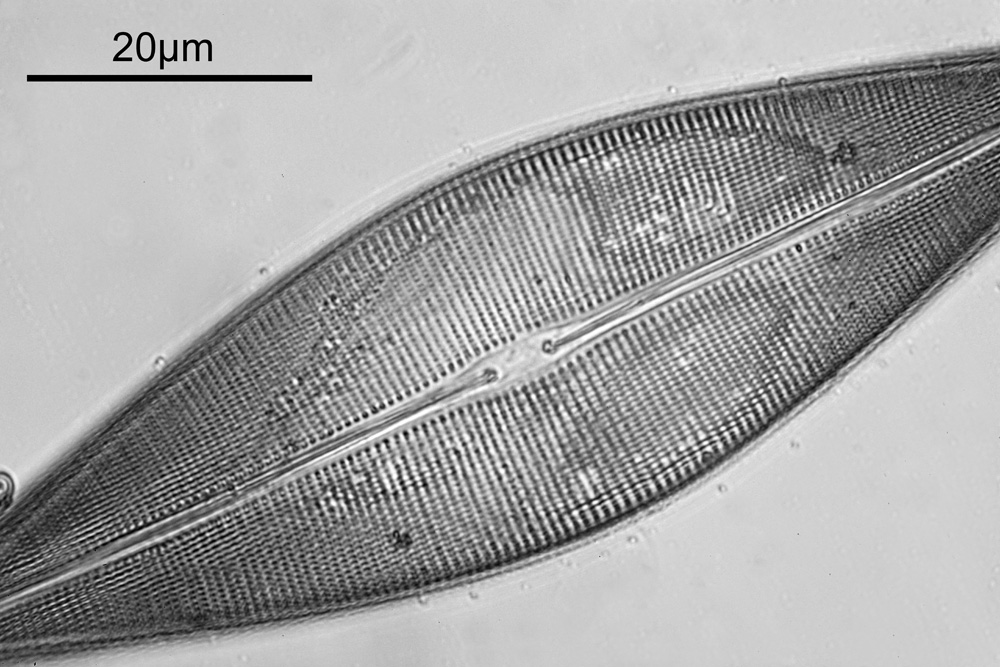
Just for comparison here’s a crop of the 63x darkfield image showing the same diatom.
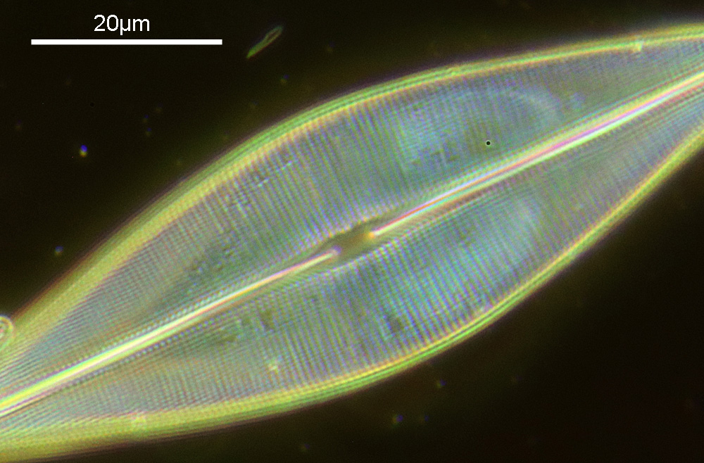
The 365nm brightfield image has slightly better resolution than the darkfield image, which is not a huge surprise – better objective with a higher NA (1.2 vs 0.9), and used a shorter wavelength of light, but some of the structures visible in the 365nm are still visible in the darkfield visible light image.
Just for giggles, here’s a crop of the 365nm darkfield image, looking at the pore structure of the diatom.
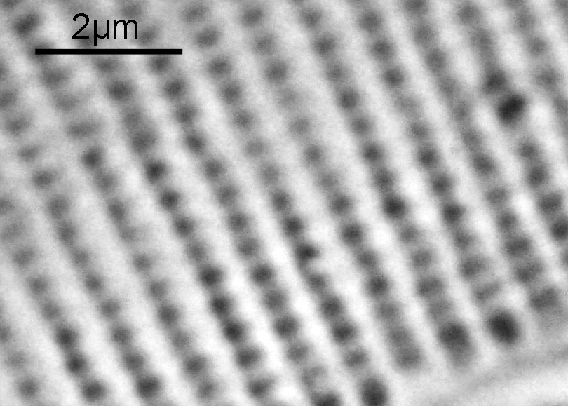
Even with 365nm it looks as though I am getting down to well below a micron in terms of the features being resolved. Keep in mind that the slide and coverslip were glass (not quartz) and the coverslip thickness was also wrong for the objective, so although this may not be as sharp as it could have been, it is still a respectable result.
It’s funny how a single historical sample can be such a fascinating thing to image. It is all too easy to forget, or just not realise, that amazing scientists existed before the days of the internet, and it can be hard to track down their work if you don’t know exactly what you are looking for. I can feel a new paper coming on looking at some of Horace Dall’s work as he was such a pioneer, and bring it to a whole new generation of scientists. If you’ve made it this far, thanks for reading, and if you’d like to know more about this or any other aspect of my work, you can reach me here.