
I recently wrote about a fascinating little microscope slide from Horace Dall, who coated diatoms in titanium dioxide to help make them more visible for microscopy (you can read about that here). Today’s post shows some new images of the diatoms on the slide, this time taken at high magnification in visible light, using a white light LED light source. I also share the objective I used for the images, as that too is an interesting piece.
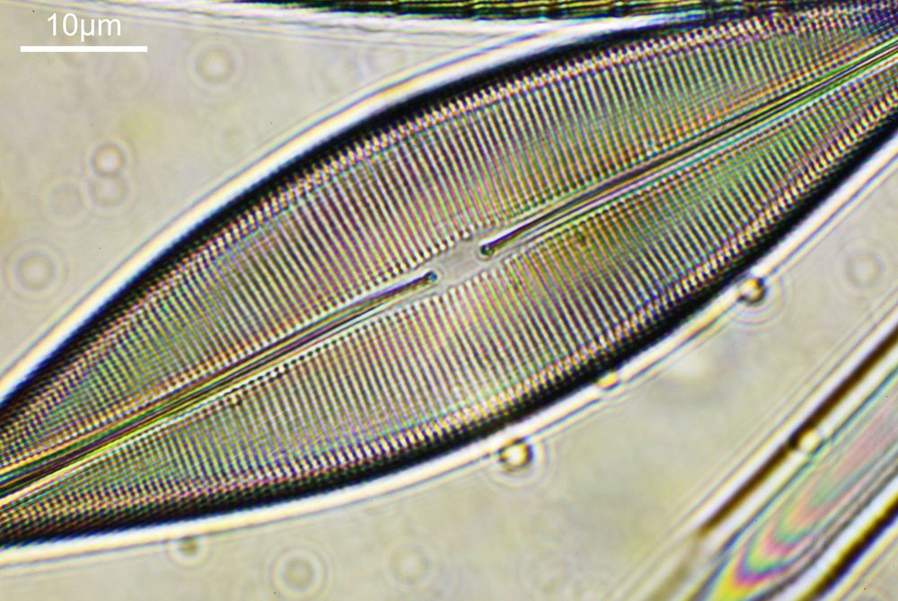
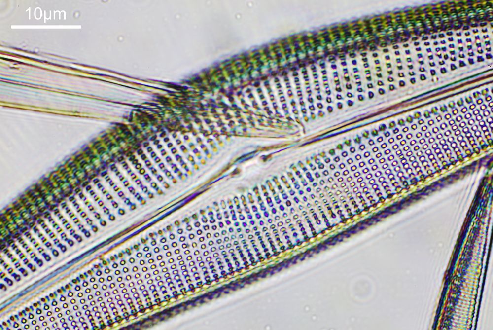
On with the images. This were taken using an unmodified Canon EOS 5DS R camera. Condenser was an Olympus Aplanat, set to about NA 1.25 and offset slightly to provide oblique lighting. This was oiled to the underside of the slide. The objective was a Leitz PL APO 100x NA 0.60 to 1.32 (wide open at NA 1.32), which was oiled to the top of the slide. Images reduced in resolution for sharing here, as the EOS 5DS R produce 50Mp images. The colours are as captured, and there is no stacking here, these are single images.
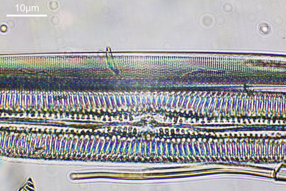
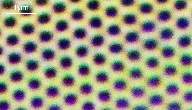
And to give you an idea of the original image resolution, here is a cropped part of the image above, shown at the original pixel resolution.
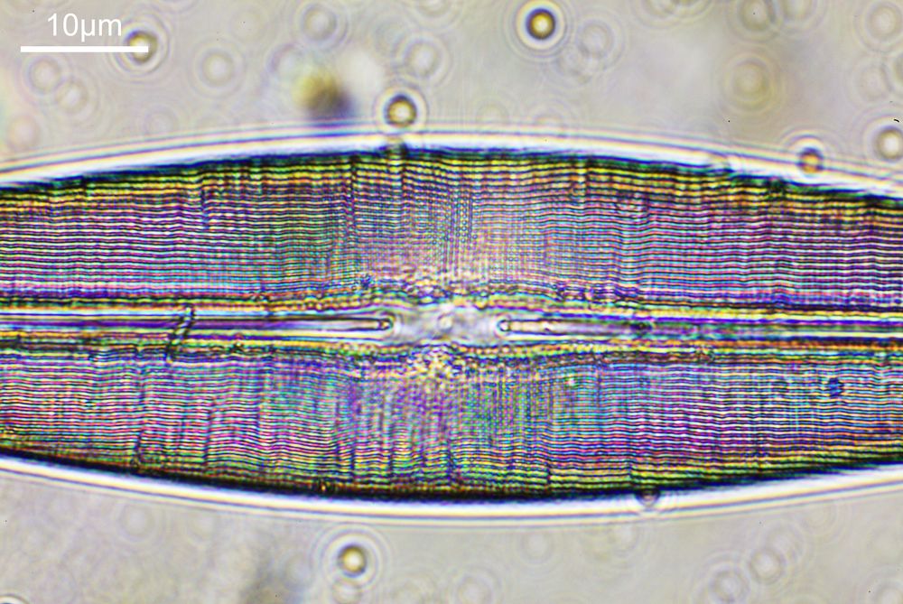
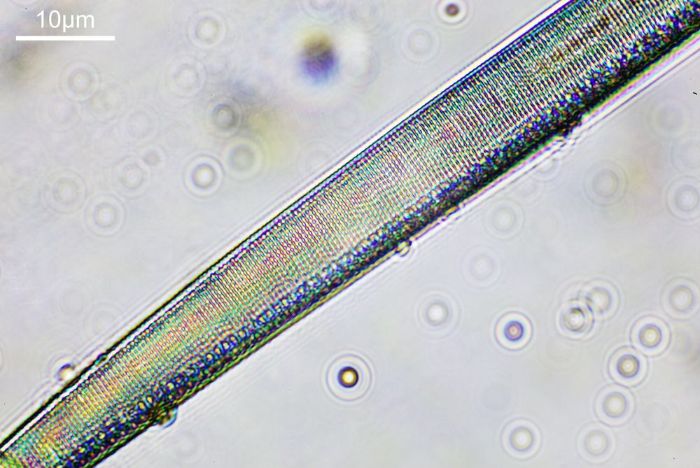
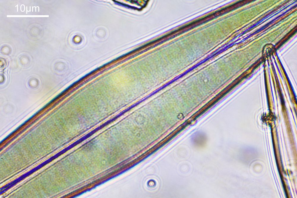
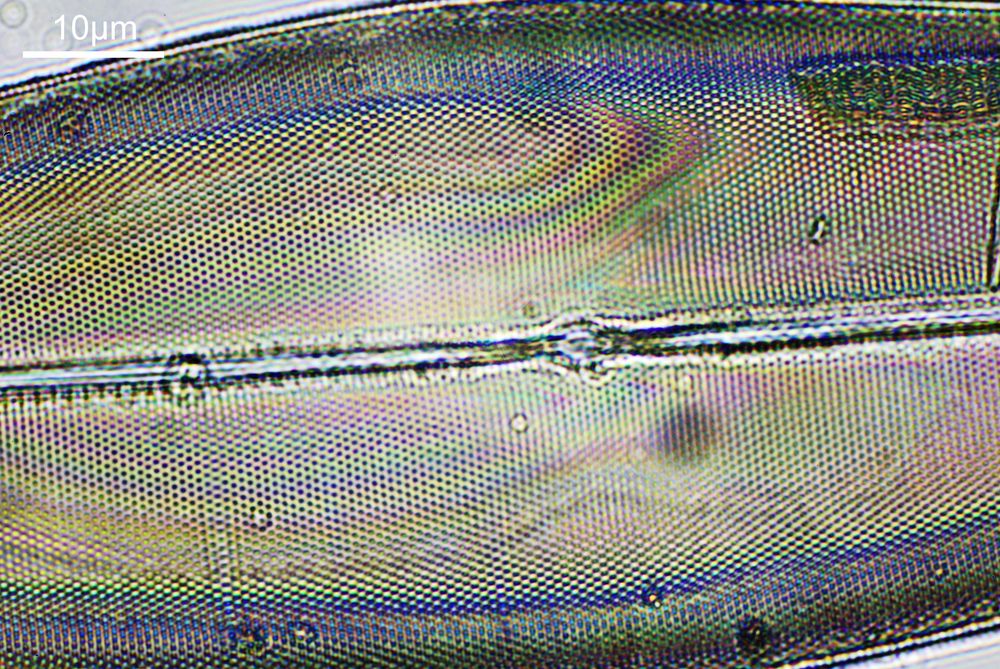
Some pretty psychedelic colours going on with the TiO2 coating of the diatoms.
As can be seen with the images the objective provided plenty of resolution, easily resolving features down to well under a micron even with using this visible light source. The objective is a Leitz 100x PL APO NA 0.60-1.32 oil immersion lens as shown below.
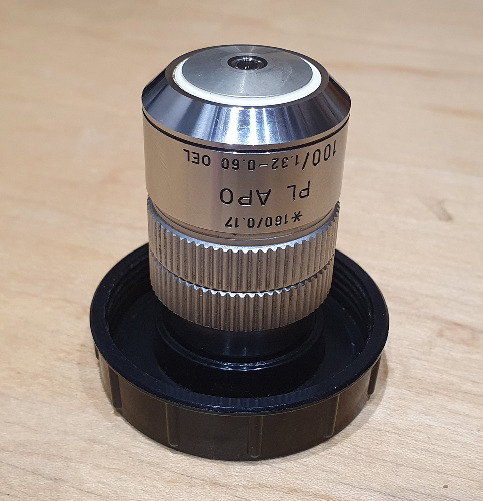
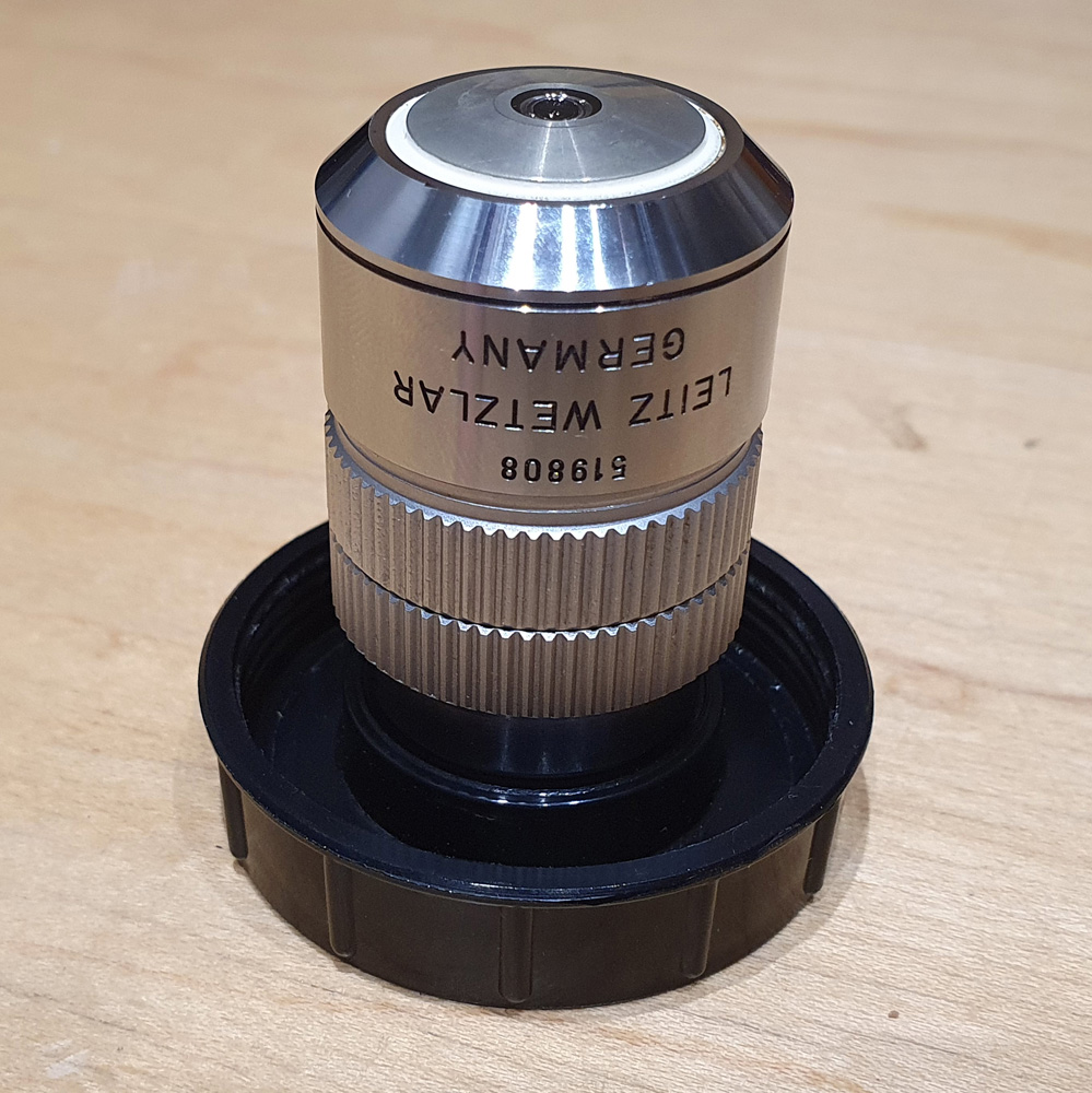
What can we tell from the name. 100 means it is 100x magnification, PL means plan (flat image) and APO means it is apochromatic, so it is designed so that three different wavelengths of light all meet at the same focal point, minimizing chromatic aberration. It is oil immersion (‘Oel’) and is for 160mm finite tube length microscopes. It also has a variable aperture, allowing it to be used from NA 0.6 to 1.32. With the images shared here it was used at NA 1.32, but being variable makes it ideal for darkfield imaging, so is a very useful feature. All in all a very capable objective.
This post is a brief one, building on previous work, so I shall leave it there for now. Thanks for reading, and if you’d like to know more about this or any other aspect of my work you can reach me here.