
Some photos are just pretty, some are challenging, others are shocking. In the world of research an image, if captured in a controlled way, can provide as much information as any other form of measurement. In some recent work, I presented imaging of a diatom at 313nm on my custom made UV microscope (see here), and one question which came from the work was how could I quantify the resolution of my objectives at different wavelengths of light, including well down into the UV. This brings me to the topic of today’s post – resolution testing of my microscope using the Newport Highres-2 high resolution microscope test slide.
To test my microscope I knew I was going to need a high end test slide – something with very small features. UV microscopy offers improved resolution compared to using visible light, and looking at my previous diatom image taken with the Leitz 100x NA1.2 UV objective at 313nm it seemed like I was resolving features down to about 200nm if not below. I also needed the slide to be made from quartz of fused silica rather than glass, so it wouldn’t block UV. As a final requirement, it needed to be able to be used with immersion oil or glycerin and a coverslip without damaging the test target. Taking all this into account I decided on a Newport Highres-2 positive slide (opaque lines on a clear background), and having spoken with them it looked like this would indeed survive being using with immersion fluid and a coverslip, and would be UV transparent. So I went ahead and ordered one.
The test slide is a USAF pattern, which has lines of different widths arranged on it. An image of the full area of the test slide taken in visible light using a 4x objective on my microscope is shown below.
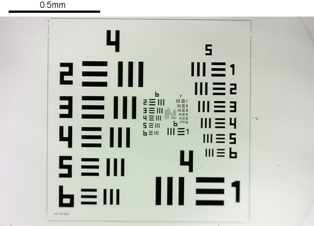
The pattern is chrome deposited onto a quartz substrate, which is mounted into the middle of a 3″ by 1″ aluminium slide as shown below. You can just about see the test pattern in the middle of the diamond shaped piece of quartz in the middle of the slide.
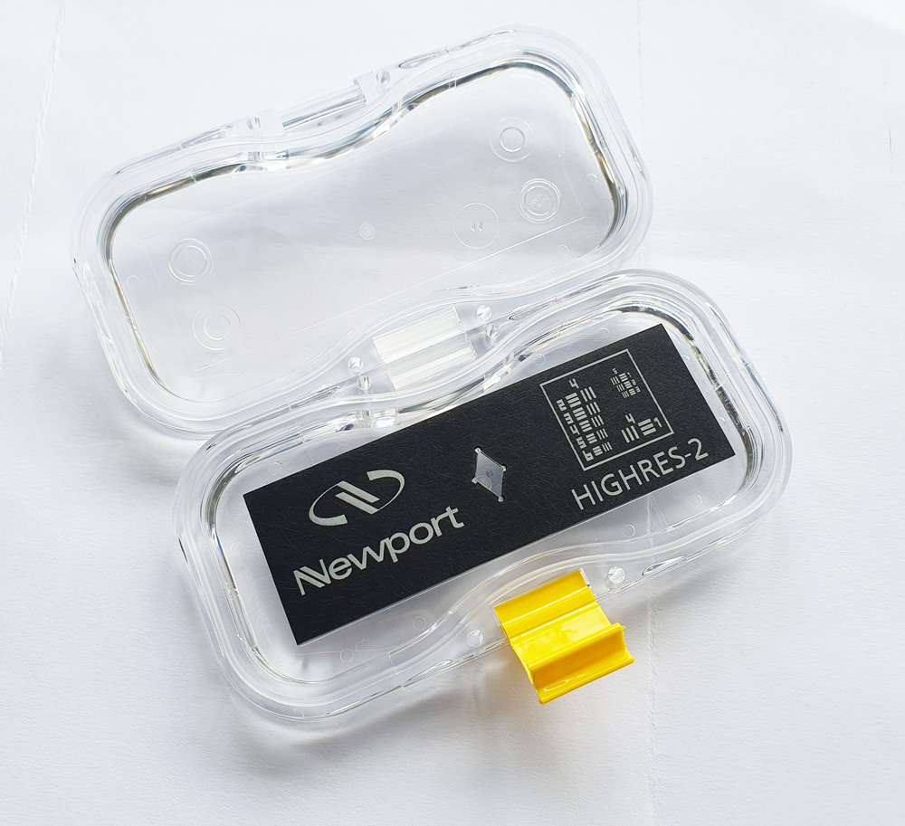
Overall it looks to be well made and with a good container which will hopefully keep it safe.
Back to the test pattern and resolution testing. The pattern is presented as a series of Groups from 4 to 11, with Group 4 having the largest lines, and Group 11 the smallest. Each group is further subdivided into 6 Elements, which again get smaller going from Element 1 to Element 6. The widths of the different lines are given on the Newport site – here.
With using the Leitz 100x NA1.2 UV objective for imaging diatoms, it looked like I was seeing features down to about 200nm if not a little smaller, so the key parts of the test slide which were of interest to me were Groups 10 and 11 as these have the smallest features. Looking with 313nm gave the following image (cropped slightly just to show Groups 10 and 11). Images were taken using a monochrome converted Nikon d800 from MaxMax, which I can use down to 300nm. I also took the image as a Raw file and processed it in Monochrome2DNG to try and get a little bit more from it as the camera sensor has had the Bayer filter removed.
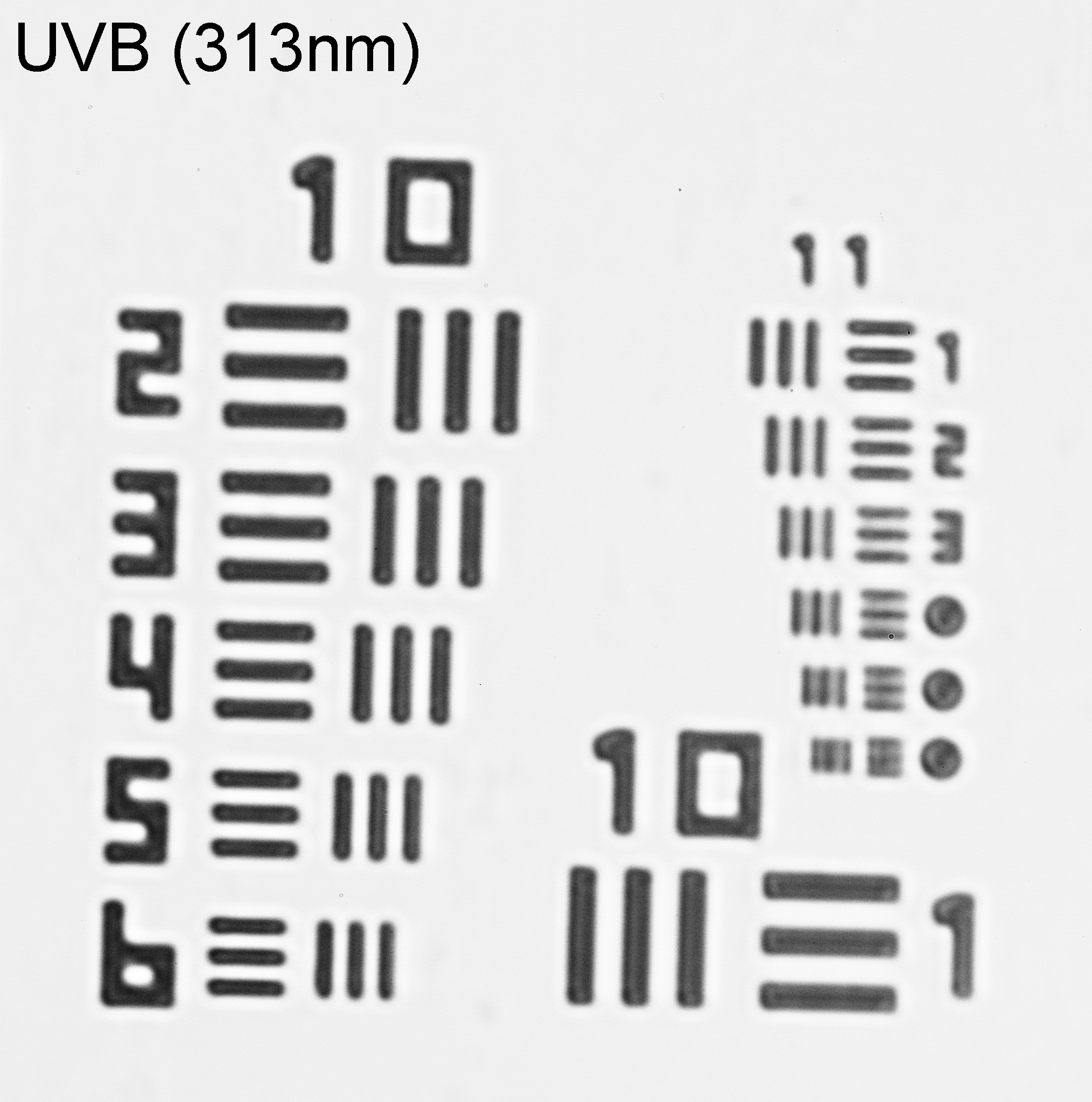
All Elements in Group 10 were clearer resolved using 313nm light. These lines go from 488nm down to 274nm wide. In Group 11, Elements 1, 2, 3 4 and 5 are clear to me, but Element 6 I’m just starting to struggle to break out the individual lines, although they can just be seen. In the image Group 11, Elements 4, 5 and 6 are identified by the dots instead of numbers. Keep in mind that Group 11, Element 6 has lines which are only 137nm across (Group 11 Element 5 is 154nm and Group 11 Element 4 is 173nm). So it does look as though that objective will certainly resolve features down to below 200nm and perhaps around 150nm. Put this into perspective – a single Covid-19 virus is about 100nm across. Not too shabby for a home built microscope.
Just for comparison here are the images taken using 365nm and visible light (546nm) captured at the same time.

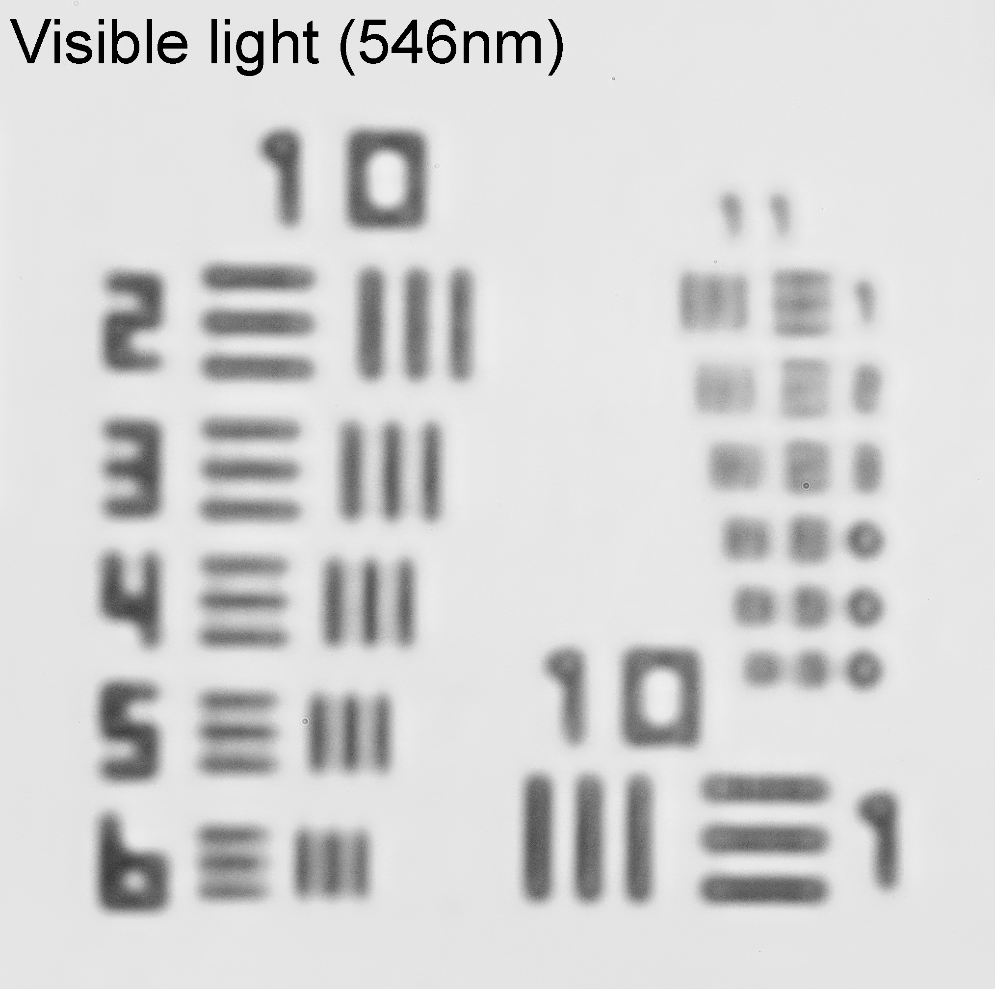
As expected, as the wavelength gets longer the resolution gets worse. Physics in action….. The quest for resolution was reason why microscopists started looking at UV transmission microscopy back in the early 1900’s. They used even shorter wavelengths of light – 254nm and 276nm – which makes me wonder how much more resolution I could get out of my microscope if I used these more extreme UV sources.
Imaging to produce nice photos is one thing, but there are times when we need to be able to put numbers against the things we image, to be able to quantify the things we are seeing. With the Newport Highres-2 resolution test slide, I have been able to do just that with my UV microscope and now have a way of being able to determine the resolution which can be achieved with different objectives and at different wavelengths.
Thanks for reading and if you’d like to know more about this or any other aspect of my work, I can be reached here.