
Coating of diatoms in a thin layer of metal or metal oxide has been reported for many years as a way of improving their visibility for microscopy (as examples, work I have discussed and published on slides from Horace Dall and John Dale. However it was never a common technique and is not widely used today. Recently I was sent a few slides from a fellow microscopist which contained diatoms which had had a thin (a few nm) coating of gold applied to them before mounting.
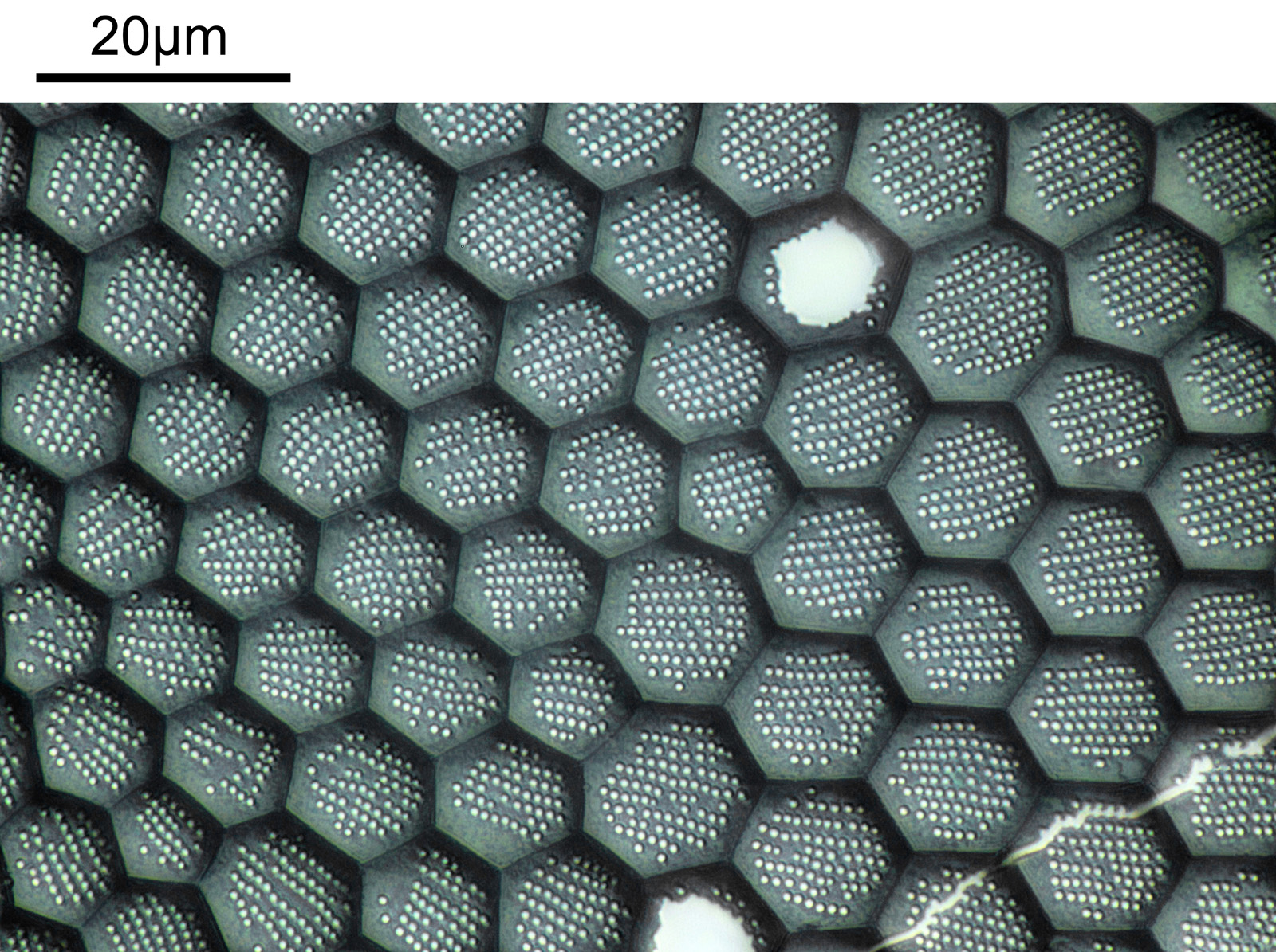
Her is an example image from one of the slides. It shows part of a diatom (likely Triceratium grande, or Triceratium favus). The resolution has been reduced for sharing here.
And the slide itself.

Imaging was done on my modified Olympus BHB microscope with a 60x Olympus Splan Apo NA 1.4 objective, and oblique illumination from below using white LED light. The image is a stack of 10 images (stacked using Zerene stacker) and photographed with a Canon R7 camera. The image shown has not been cropped – this was the full field of view.
Gold coating of of the diatoms certainly improves the visibility of the features, and I am looking forward to looking at the other samples. Thanks for reading, and if you’d like to know more about my microscopy or other aspects of my work, I can be reached here.