
This post finishes off something I started yesterday (see here) where I tried pushing the resolution I managed to get on a couple of diatom slides while only using 450nm light. The N.B.S. slides I imaged had diatoms with small features on them, but one really difficult diatom to photograph using visible light is Amphipleura pellucida, as this has features down around 250nm in size. While I have done some work with this using UV light (for example here), my challenge was to try using 450nm blue light in combination with circular oblique illumination and even cross polarization.
Equipment wise I used my Olympus BHB microscope with the Reichert Neo condenser, in combination with a 100x Leitz Pl Apo NA 0.60-1.32 objective. Kept wide open, this objective has such a high NA, the result is that instead of dark ground, I get a circular oblique illumination. 450nm LED light. No stacking was done, but for the non-polarized image I did try averaging 10 images in camera. Cross polarization was done by adding one linear polarizer just above the field lens, and another on top of the photoeyepiece, rotating the bottom one until the image was at its darkest. The images showing the full diatom have been reduced in size for sharing here, but I’ll also include crops at original resolution to better show the features.
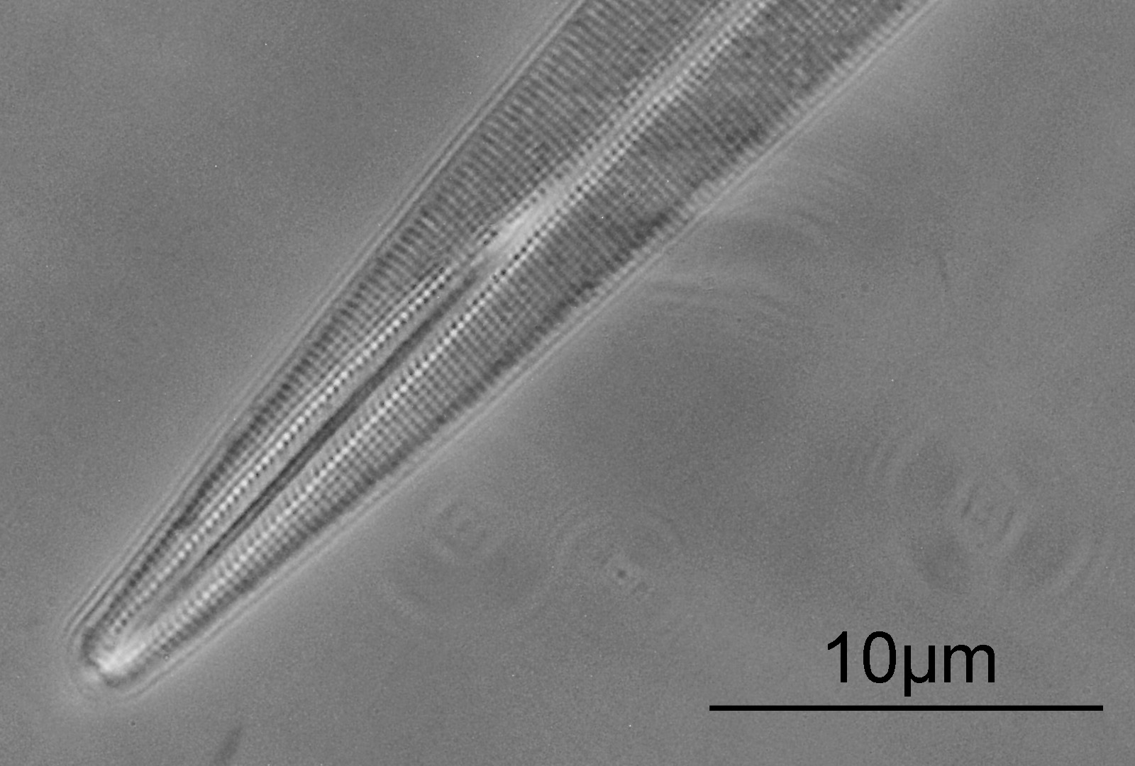
First the non-polarized image, so just using circular oblique illumination.
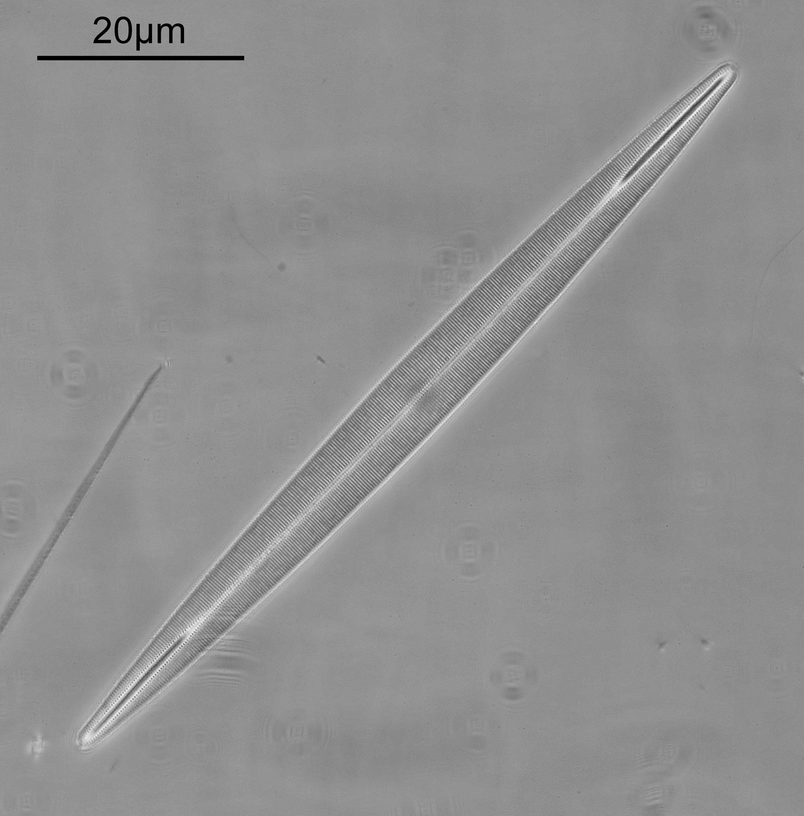
The striae are nice and obvious with this setup, and can be more easily seen by looking at a closeup of the top right of the diatom.
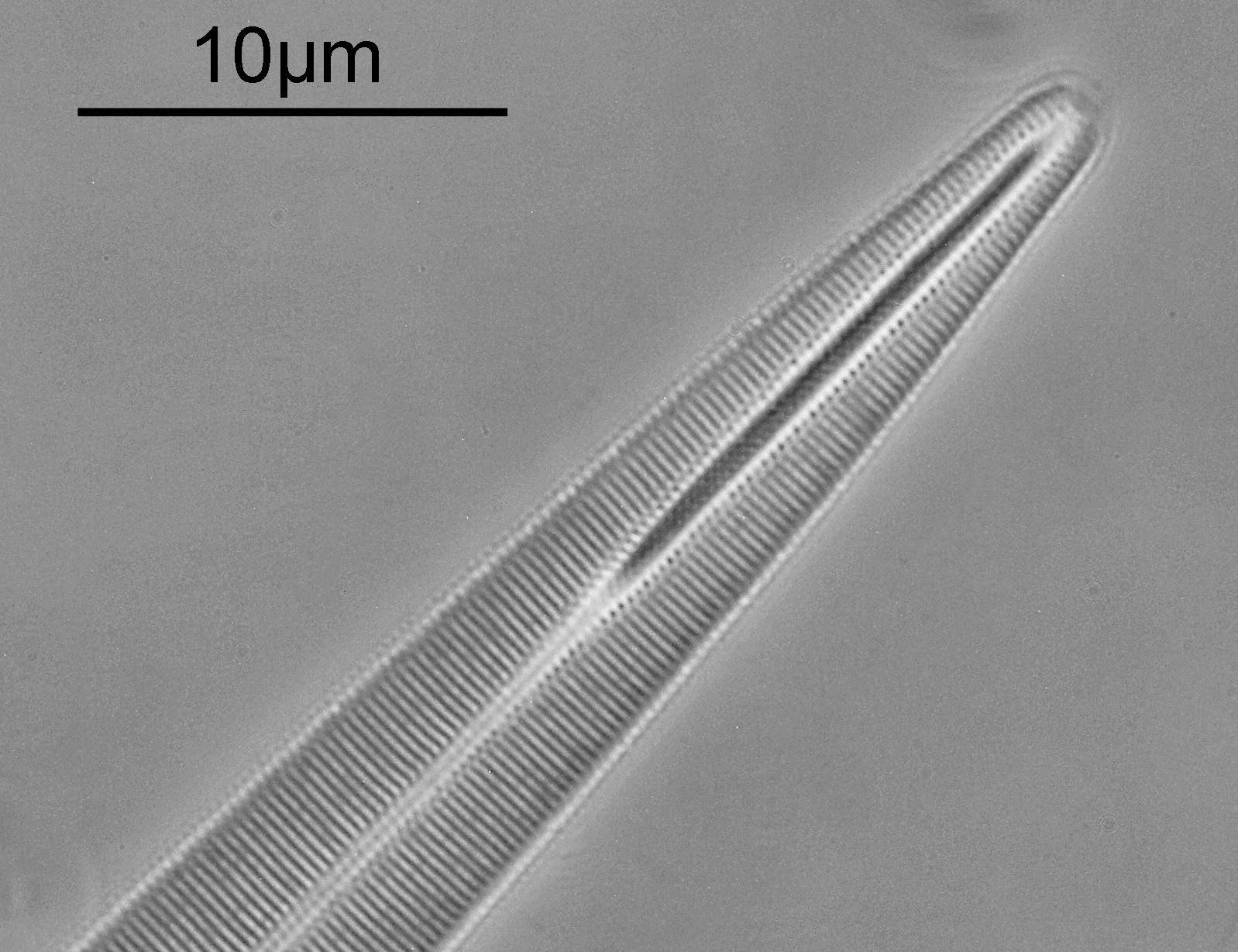
Putting this into ImageJ and doing some measurements, we have 10 striae in 2.659µm (or 37.6 striae per 10µm). This matches pretty well with the slide which says 40 lines per 10µm. Think of it another way, those lines in the image above are 266nm apart.
Cross polarizing in combination with oblique lighting is well known as a way of showing the punctae on this diatom, so I tried that approach. I placed a linear polarizer on the field lens (below the condenser), and then another on top of the photoeyepiece. I rotated the one on the field lens until the image was at its darkest which would be when the polarizers are aligned at 90 degrees to each other, and then took an image of the diatom.
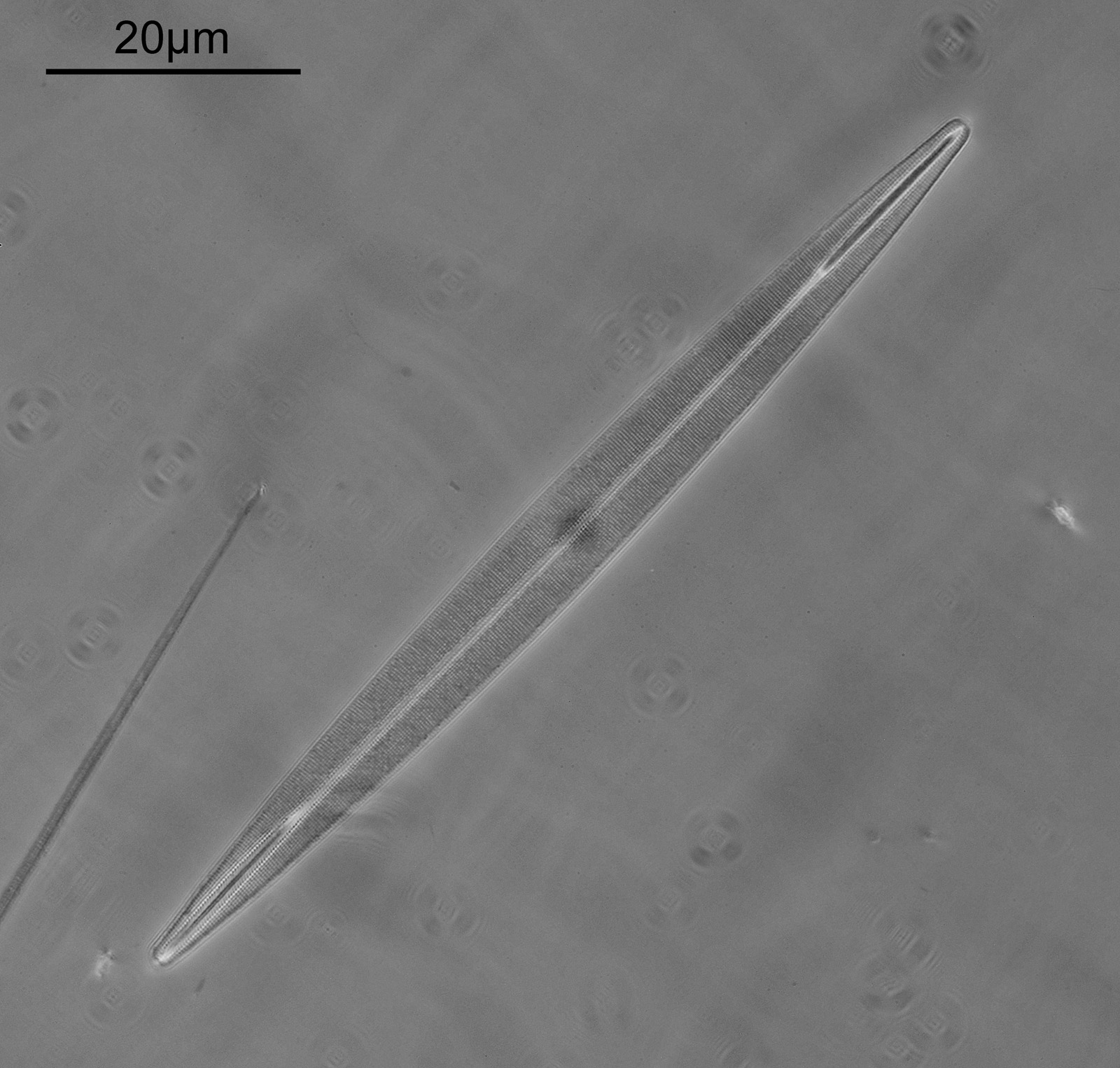
Even looking at the whole diatom, the lines appear a bit more broken up, but this can more easily be seen on closeups of the top right and bottom left of it.
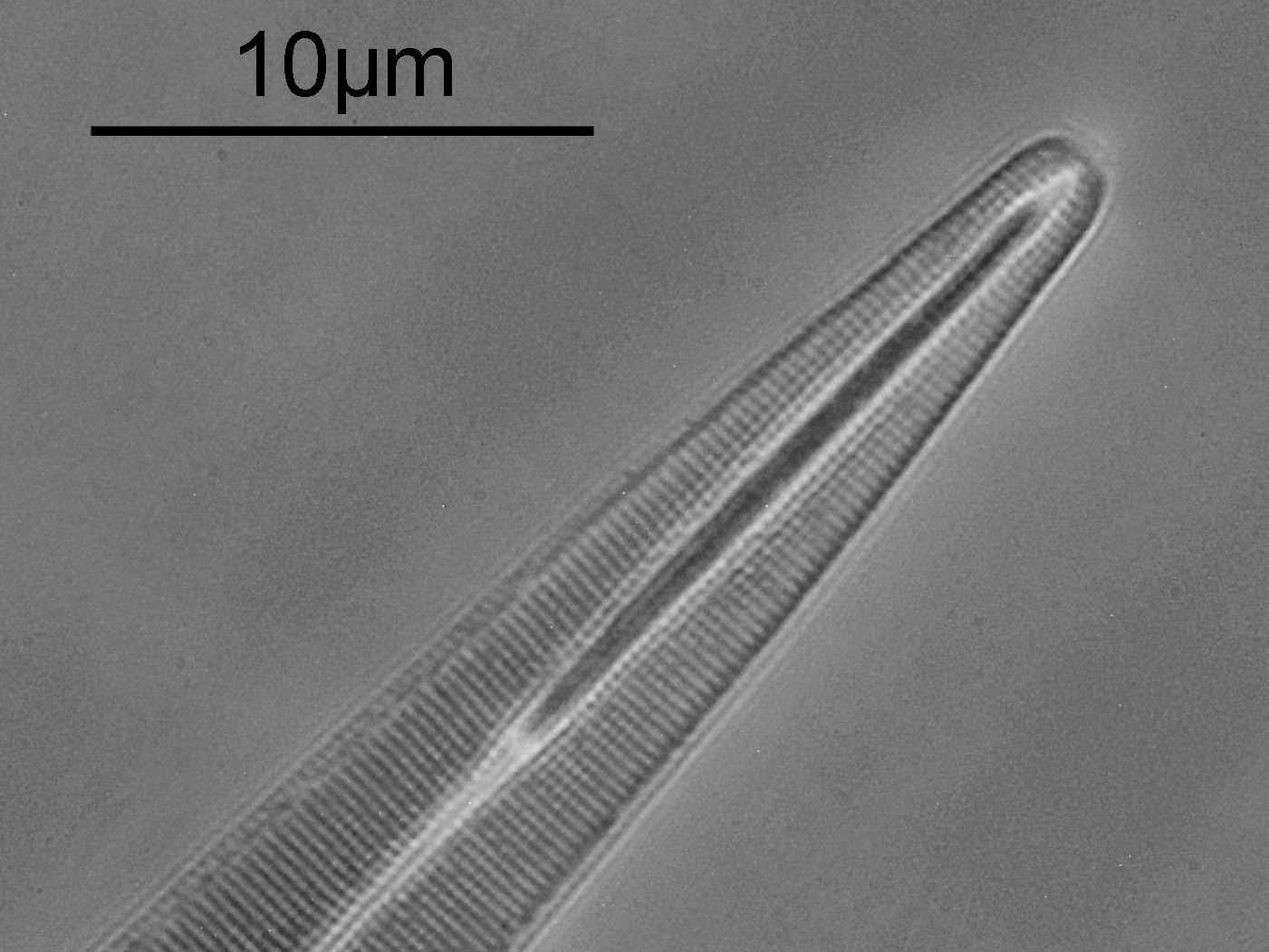
With cross polarization, I’m just starting to see a hint of punctae of the diatom (dotting), where the lines are breaking up into individual features, so has been beneficial for the imaging here. For using 450nm light I am happy with this result, especially at the first attempt.
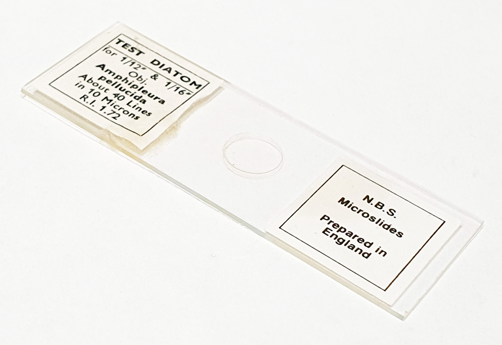
Before I go, here’s the slide.
As always, thanks for reading, and if you’d like to know more about my work, I can be reached here.