
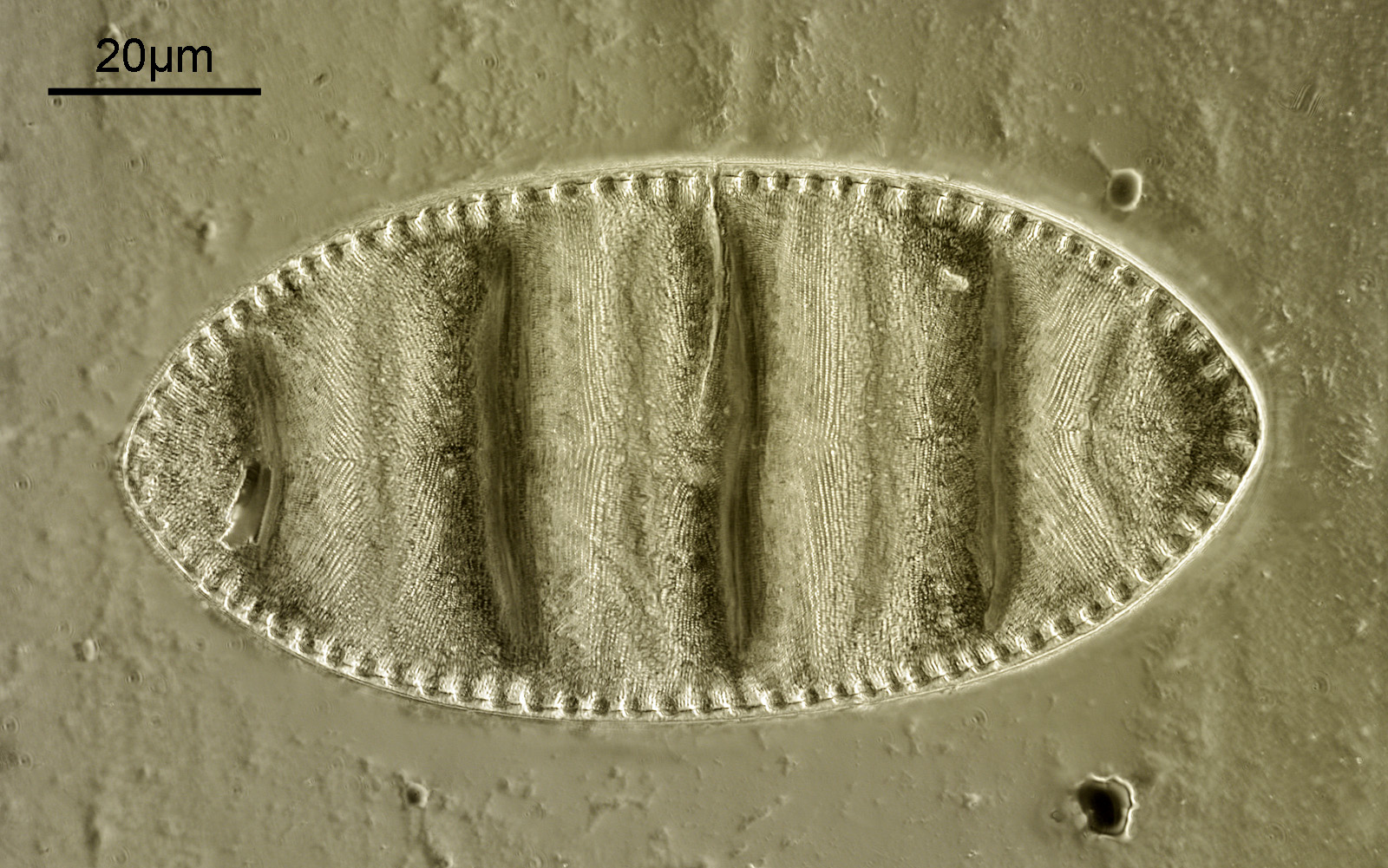
A few days ago I took a look at a Klaus Kemp slide of diatoms called Cymatopleura elliptica with 450nm light and a Leitz 63x Pl Apo NA 1.4 objective and, well, the image was not as good as I had hoped. Today I decided to revisit the slide with a different approach, and this post shows the results of that work. The image was done on my modified Olympus BHB microscope, light source was a Zeiss Mercury Xenon lamp. Objective was a Leitz 100x Pl Apo NA 1.32-0.60 (oil immersion). Condenser was a Watson Holoscopic one (again with oil). Objective iris was closed down slightly with the aim of trying to produce circular oblique lighting (COL). Photoeyepiece was a Nikon 5x CF. Filters – 365nm (10nm bandpass, Edmund Optics, 2 stacked together), 405nm (10nm bandpass) and 450nm (40nm bandpass) from Thorlabs. Camera – monochrome converted Nikon d800 from MaxMax. At 365nm, a stack of 18 images processed in Zerene stacker. Image cropped slightly and reduced in resolution for sharing. The camera produces slight colour casts and I have kept those here as I like the effect. And after all that, here it is.
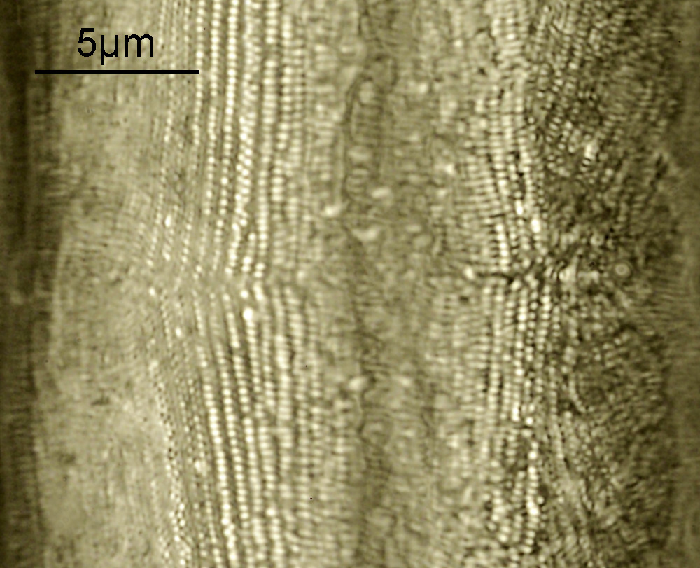
Moving to 365nm light produced a noticeable improvement in resolution, although that is probably lost a little on the image above which is 1600 pixels across compared with the original (5826 pixels across). As an example of the resolution of the original, the image below is a crop before it was reduced in size.
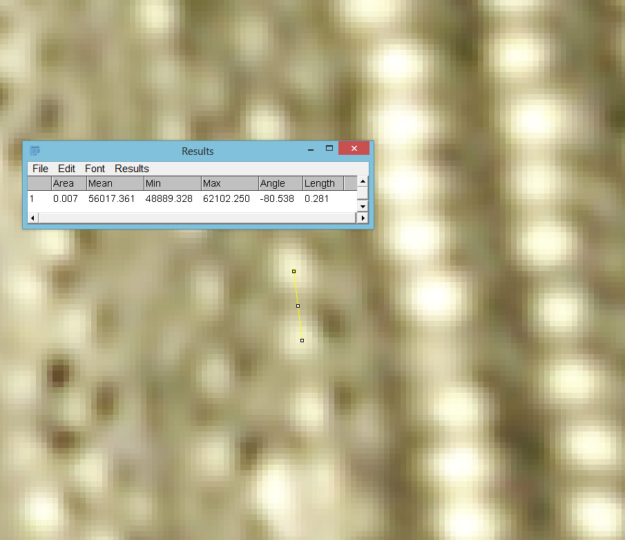
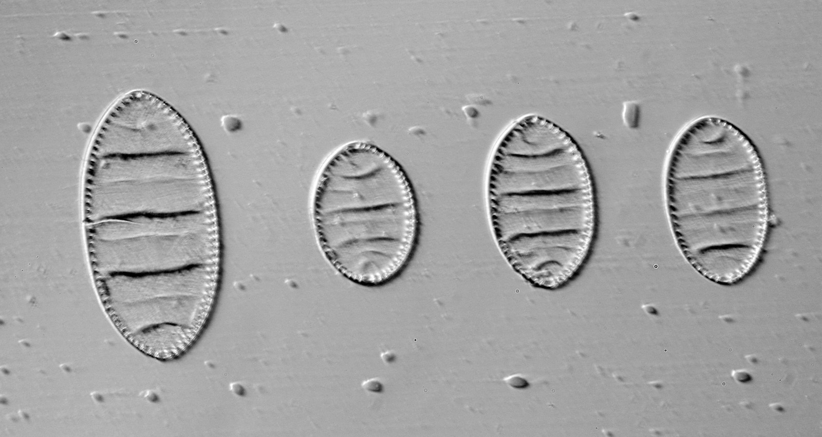
There is plenty of detail in the image, much more than my initial attempt with 450nm light. Going in closer and checking in ImageJ, some of those features are about 280nm apart.
While the slide was on the microscope I took a series of 3 images of it at 3 different wavelengths (365nm, 405nm and 450nm). These are shown below.
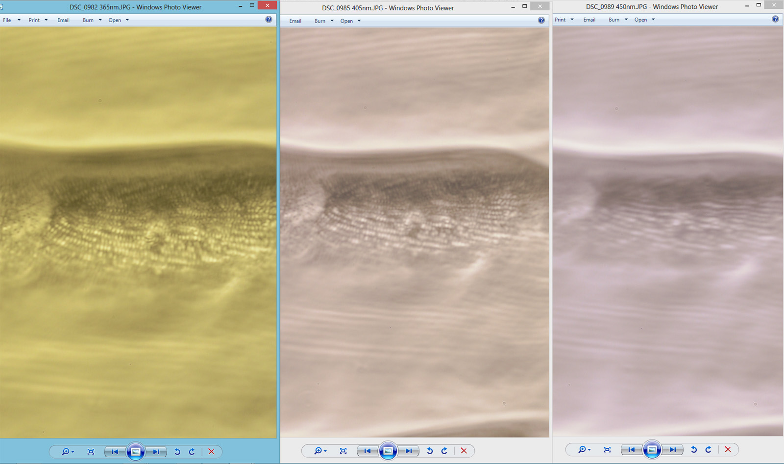
The images above are unprocessed and not sharpened, but the microscope was refocused each time. It demonstrates nicely how the shorter wavelength light brings out the features in more detail (as expected by Abbe).
The slide actually has 4 examples of the diatom on it. The larger one was the one that was imaged.
It looks as though there may something unusual with the fixative, as shown by the droplets in the image which were very visible on this slide. These were not on the outside of the slide or coverslip. Here’s the slide.
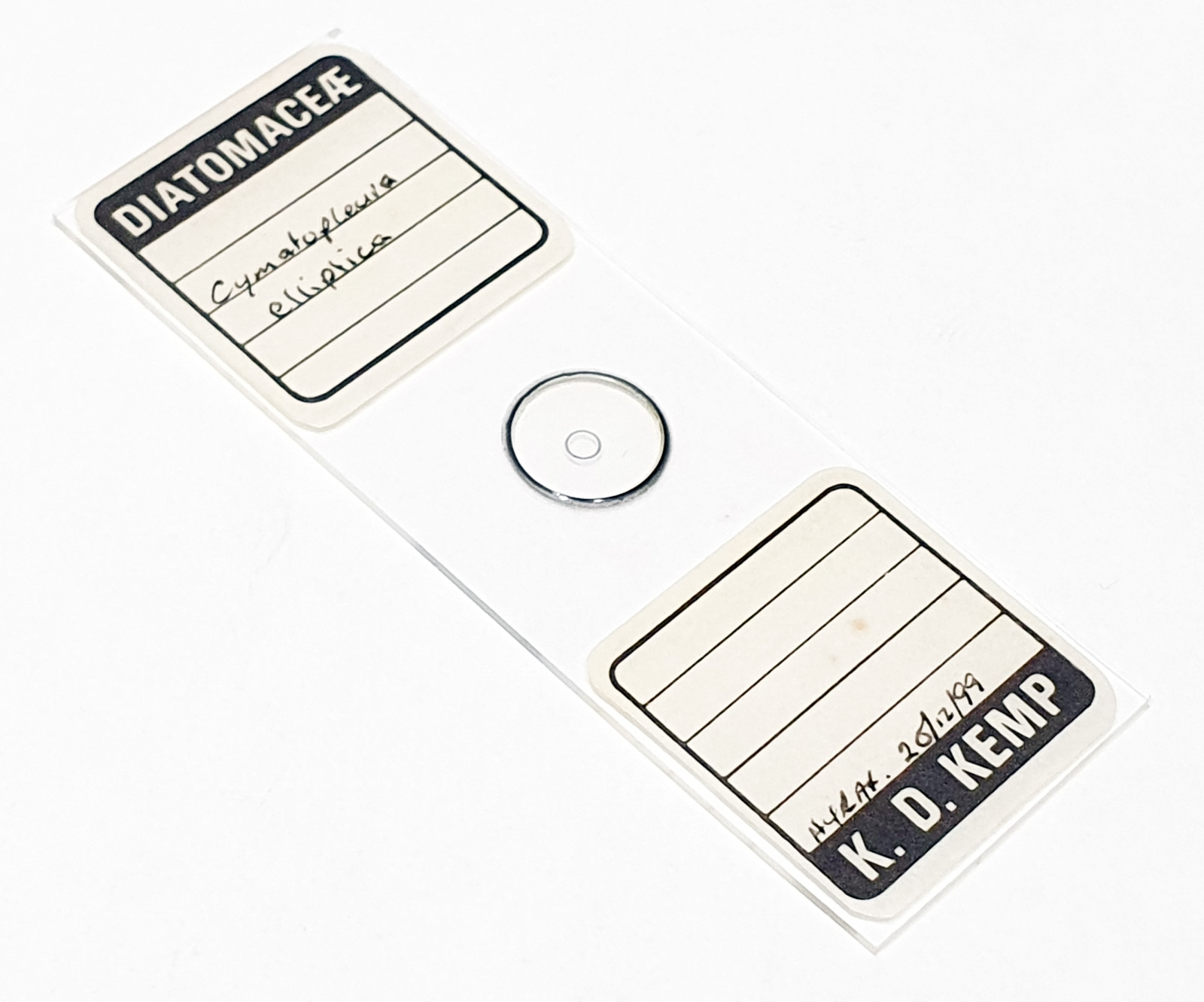
The mountant is Hyrax which was one reason I decided to try 365nm light (it has pretty good UV transmission at 365nm). What was more surprising was the objective – I expected the Leitz 100x Pl Apo to block a lot of the UV and it actually performed really well. I must remember this for future work. Diatom naming is still well beyond me, but this one is also sometimes referred to as Surirella undulata (see here).
UV microscopy presents its fair share of challenges, but imaging at 365nm is the more straightforward end of the spectrum to try and provides some quite visible improvement in resolution compared with visible light. Be safe though if you plan on trying this – I use UV safety specs whenever dealing with UV light sources. As always, thanks for reading, and if you’d like to know more about my work I can be reached here.