
Melanin acts a primary skin defense against UV, helping to absorb it before it can reach too far into the skin. I was intrigued by what skin with a high melanin content would like like under the microscope and recently managed to find a prepared slide of highly pigmented skin (I’m guessing Fitzpatrick V or VI) and wanted to share some images, as they nicely show the melanin.
These were taken on my Olympus BHB (affectionately named Project Beater, given the state it was in when I bought it). The light source was just the standard tungsten filament bulb, and the images are all bright field. Objectives between 4x and 63x were used, and approximate scale bars included in the images (which are just single shots, no focus stacking). Camera was a Nikon d850 monochrome conversion, with no filtration. Here are the images;
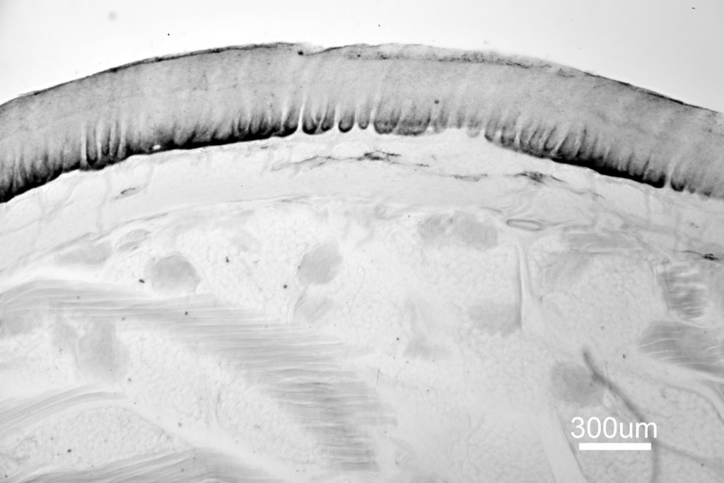
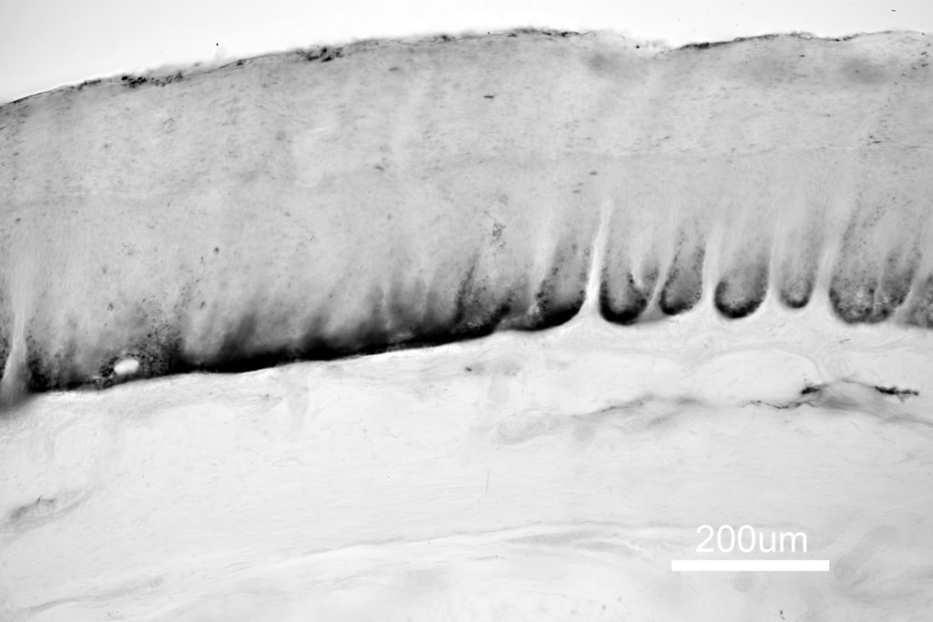
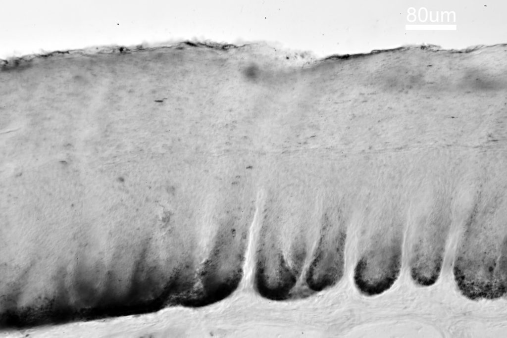
As can be seen in the 4x image, the skin sample includes the stratum corneum (top on the image), and the viable epidermis, the dermis and the subcutaneous tissue. At the bottom of the viable epidermis, where it interfaces with the dermis, there is a really dark layer. This is where the melanocytes are present, and they produce the melanosomes (the pigmented melanin granules). These melasomes are then transferred into the keratinocytes, which through the process of division and differentiation are pushed upwards and eventually form the corneocytes of the Stratum Corneum, taking the melanin with them.
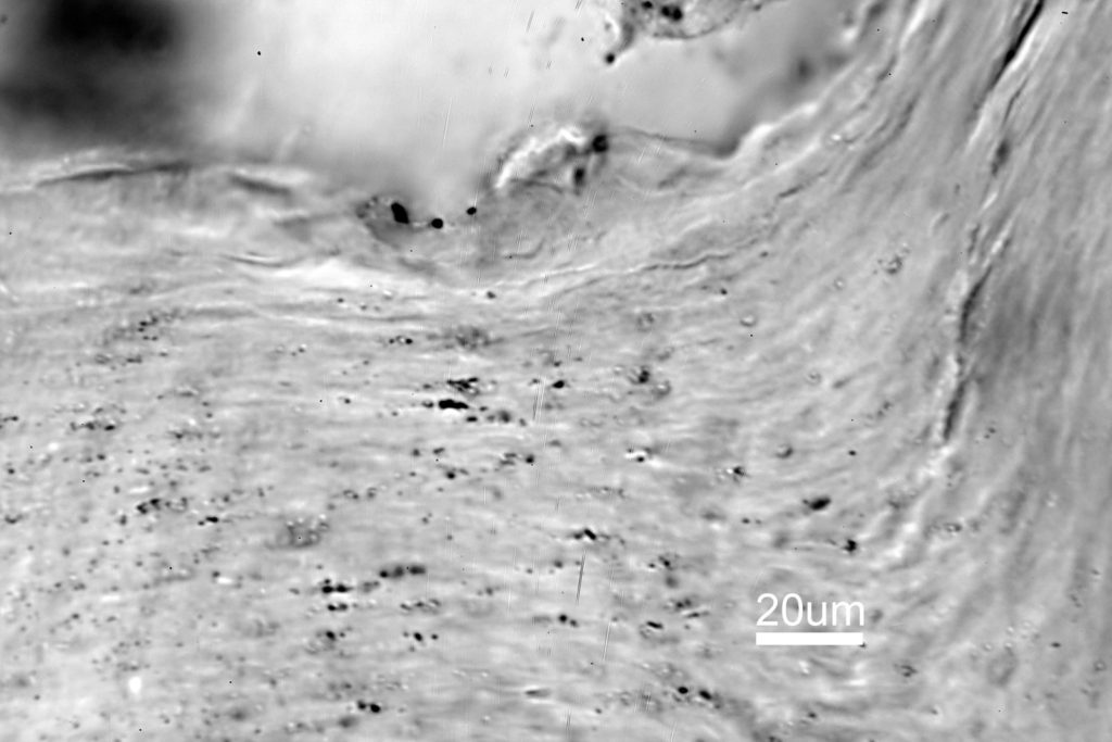
In the image with the 63x objective we are now looking at the top layer of the skin, the flattened corneocytes of the Stratum Corneum. At this magnification you can see individual and clusters of melanosomes, which appear as black dots in the image. Amazingly these are sub micro in size – literature gives sizes of a few hundred nanometers across for them.
The images here were taken with normal visible light lighting. It’ll be interesting to come back to this slide at some point with UV imaging as melanin absorbs more light at shorter wavelengths. I’d also like to try some higher magnification images with focus stacking to see if there is more detail to be had with the melanin granules.
Microscopy opens a window into worlds we cannot see with the naked eye, and the more I research it the more I am fascinated by the images it can produce. Thanks for reading, and if you want to know more about this or other aspects of my work, I can be reached here.