
This post is a bit of a double whammy. First, I’ll share some diatom images from a microscope slide with a very unusual mountant – mercury iodide. Second, I’ll talk about the useful and multifunctional objective I used to take these images – a Zeiss 40x Plan Apo, with an iris and a phase contrast ring.
Enough talk. Images. Here are 4 images of a diatom called Pleurosigma angulatum from the slide by JD Möller. Imaging was done on my modified Olympus BHB microscope, using 450nm LED light, and with a monochrome converted Nikon d850 camera.
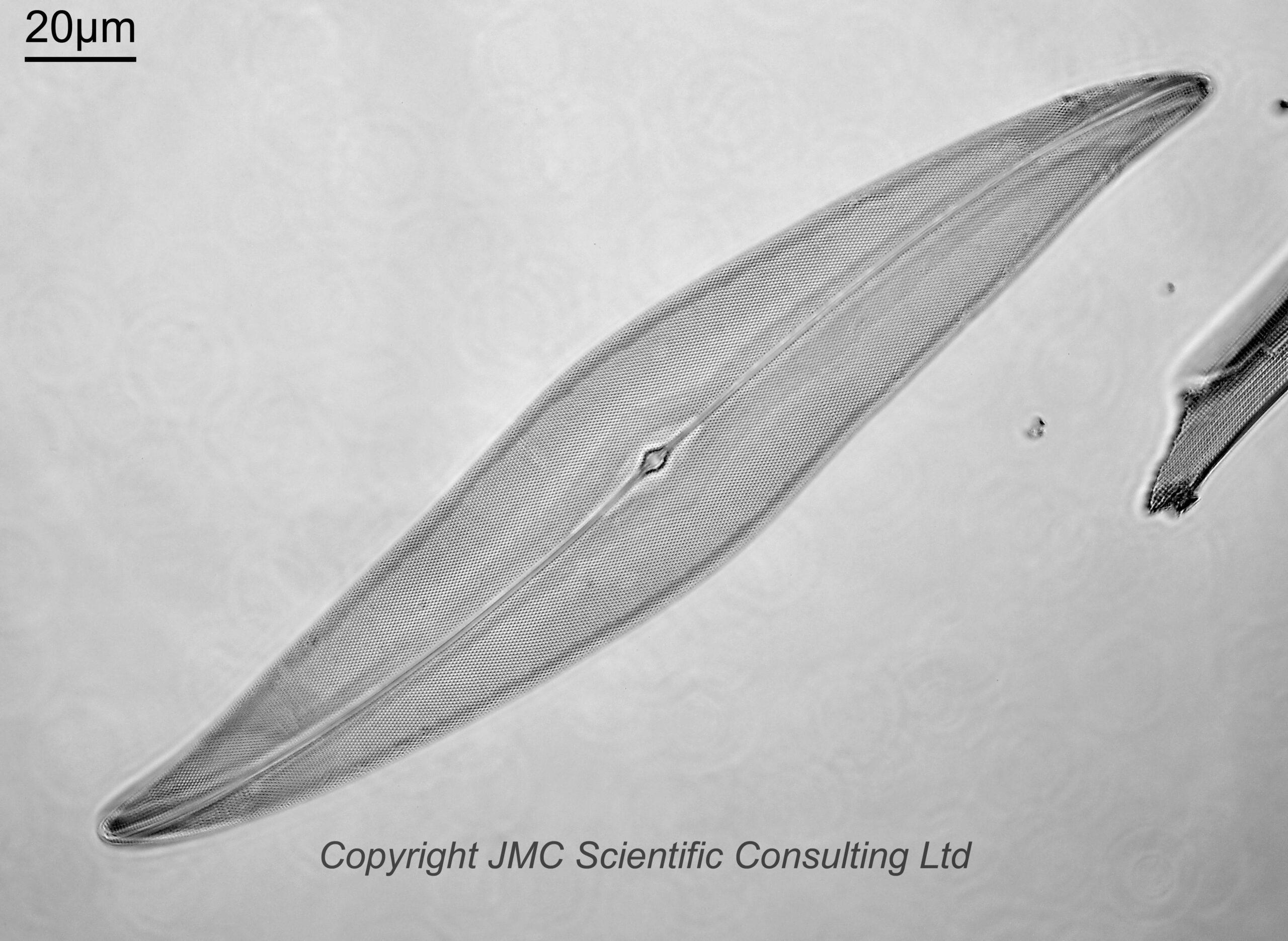
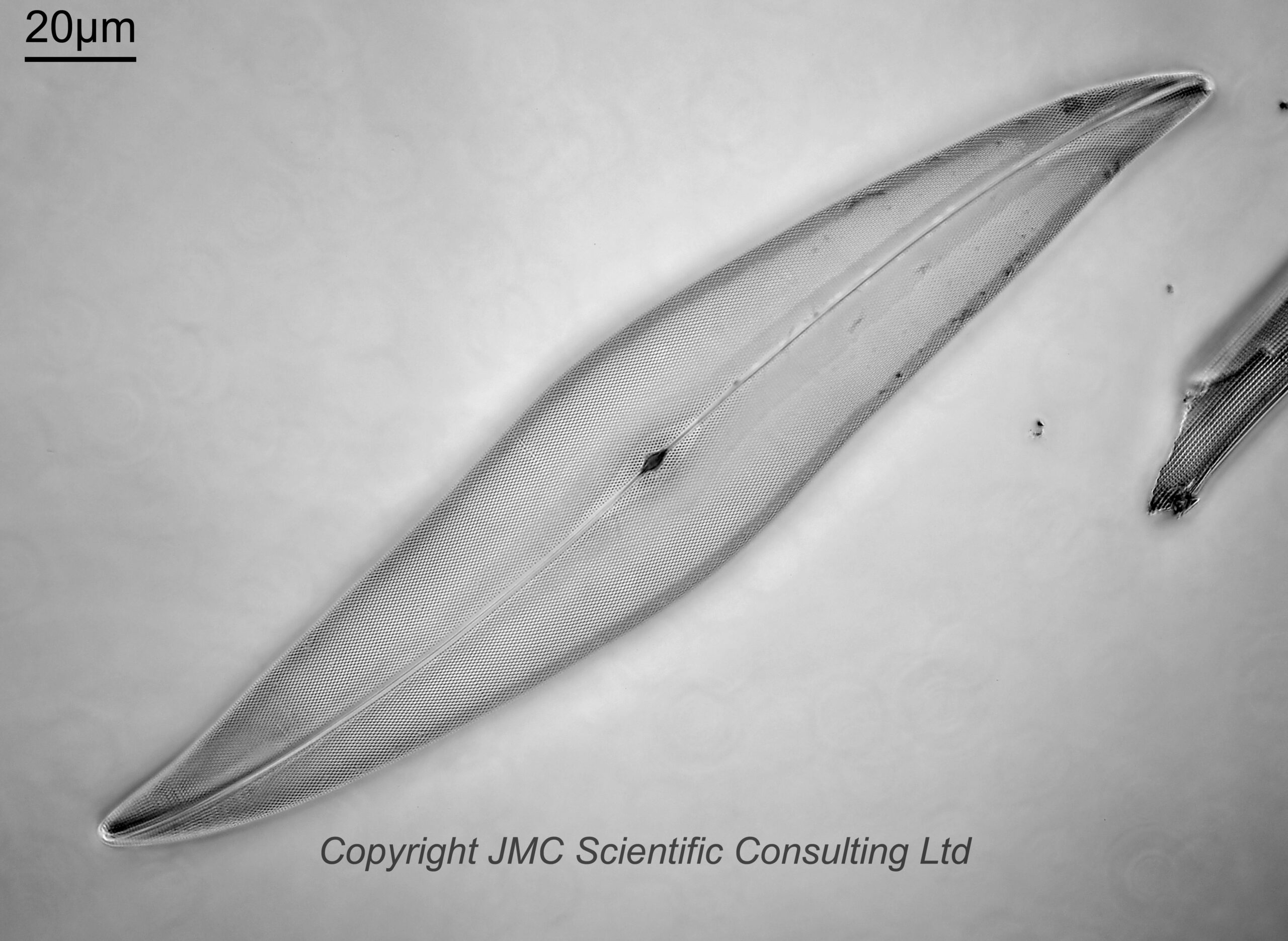
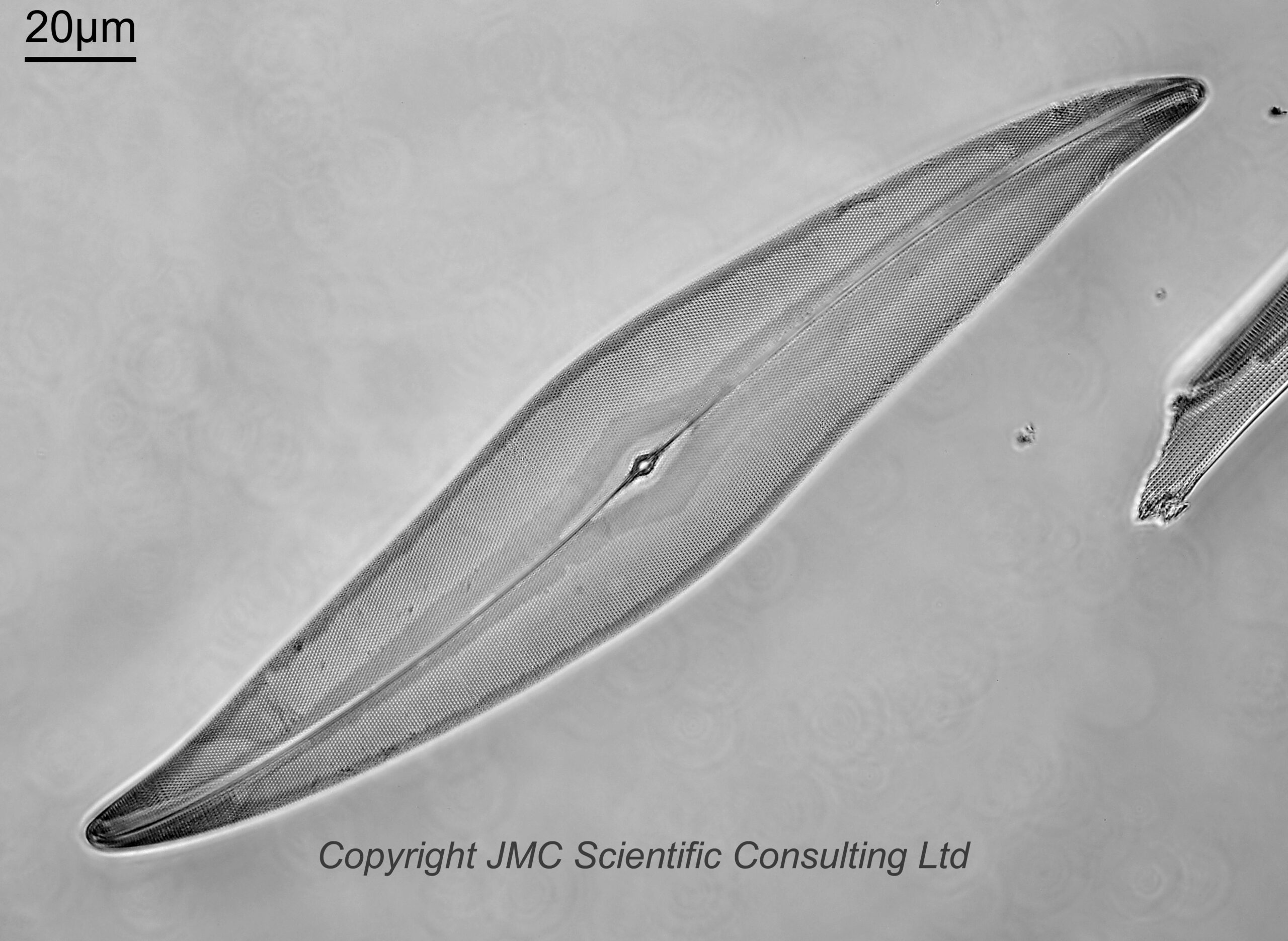
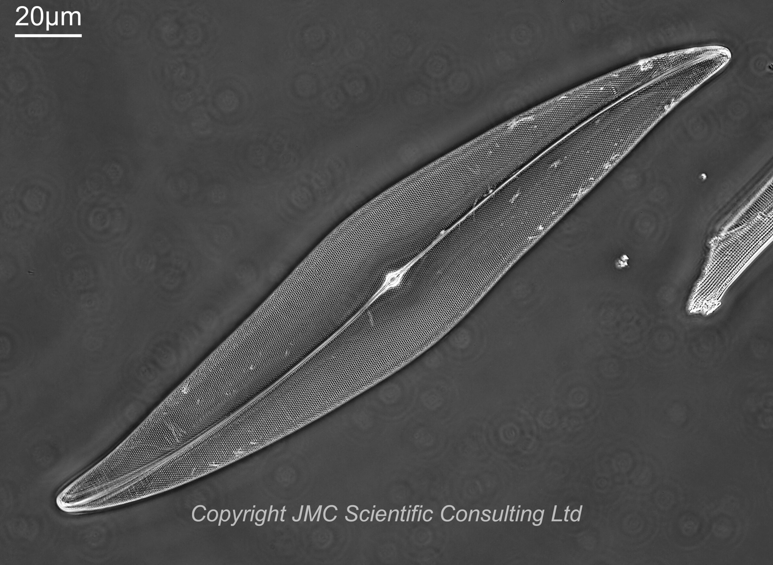
For these images I used a Leitz Heine condenser (this was not oiled to the underside of the lens, and was used without the top oil lens attached). The first two images were done with the condenser set to brightfield illumination, and with the iris on the objective fully open, and then slightly closed down. For the next image, the iris on the objective was opened back up, and the Heine condenser position moved to between brightfield and darkfield. This is supposed a phase contrast setting, and the image does look to be (at least partly) a phase one to me. Then for the final image the Heine condenser was moved to the darkfield position, and the iris on the objective left wide open. This a darkfield/circular oblique lighting image. This was where I must admit to a bit of a mistake. I should have closed the iris on the objective down a bit to make it more darkfield. Oh well, c’est la vie.
The images all show the dot pattern on the diatom nicely, but with variations in contrast between different areas, and have their own aesthetic qualities.
The Leitz Heine condenser was an interesting one to use for this, as by moving it up and down you can go from brightfield, through phase contrast, and to darkfield all with one condenser.
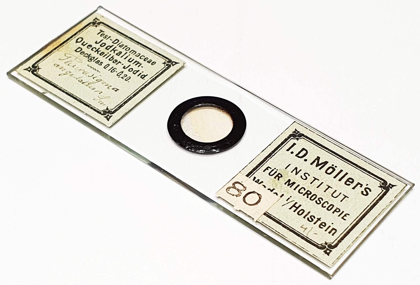
The diatoms were on a slide by JD Möller, and used the mountant ‘Jodkalium-Quecksilber-Jodid’, which is Potassium Iodide/Mercury Iodide. This is a high refractive index mount, with an RI of around 1.75-1.80 based on the information I have been able to track down. This is much higher than Styrax which was commonly used at the time, and this comes from an era before the likes of Naphrax, when a lot of experimentation was being done to find high RI materials for mounting diatoms for microscopy. Here’s the slide.
Now, the second part of this post – the objective that was used. This is a 40x Zeiss Plan Apo with a phase ring and an iris.
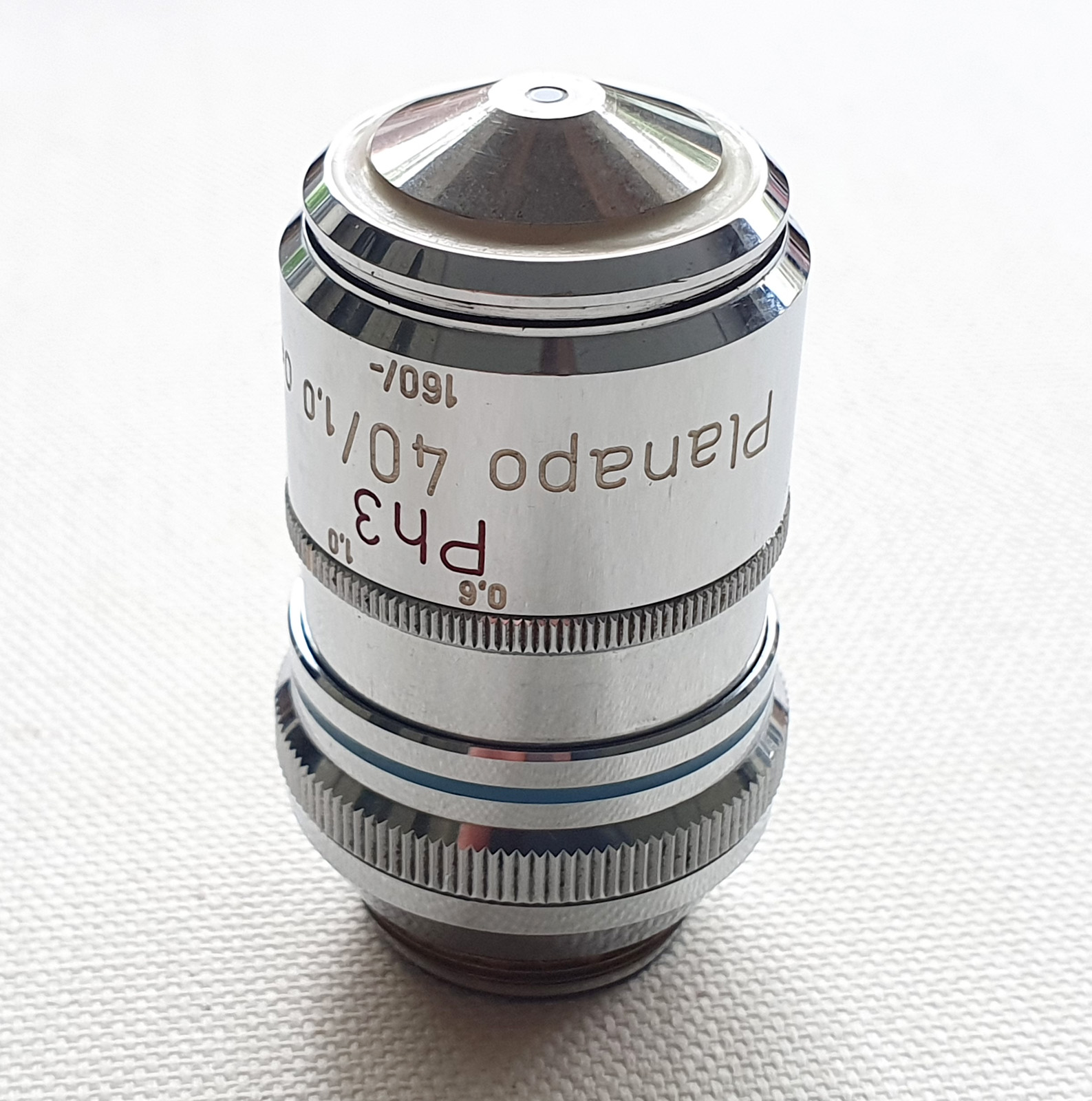
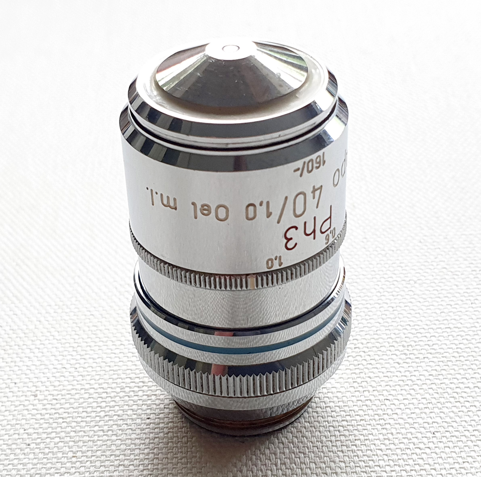
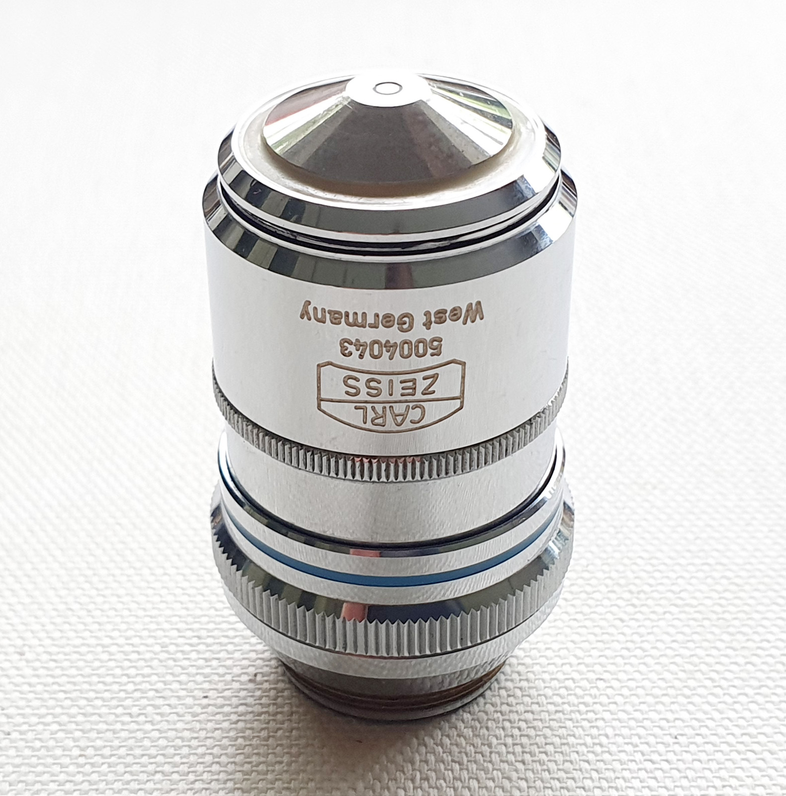
There’s a bit to unpack with this lens, so I’ll take you through the labels. This is a 40x objective. It is a high quality Plan Apo one. Plan means it is a flat imaging field, and Apo meaning apochromatic (corrected for chromatic aberrations across a wide spectral range). Plan Apo typically offer the highest optical quality from the range of lenses provided by a manufacturer. It has an an NA of 1.0, and is designed for use with oil immersion (Oel). It has an iris (m.I.) which can vary the NA between 1.0 and 0.6. Varying the iris can be useful when for example doing darkfield imaging. It it is also a Phase Contrast objective (Ph3) and has a phase ring if you look down it from the back. It is for 160mm tube length microscopes and can be used on slides with or without a coverslip (the ‘-‘ in the 160/- on the microscope barrel). All in all a very, very, useful and multifunctional objective, and would no doubt have been quite expensive when new.
As mentioned above, for these images I used the objective in combination with a Leitz Heine condenser which can be moved to provide brightfield, phase contrast and darkfield illumination all from the one package. This condenser and objective used together provide a lot of options for tweaking the image to give the desired final look.
As always, thanks for stopping by, and if you’d like to know more about this or other aspects of my work, I can be reached here.