
I built my UV microscope to help with my sunscreen research, but in doing so I quickly realized that UV offered the chance of improved resolution due to the shorter wavelength of light being used. While not so important for looking at sunscreen emulsions, this has proved very interesting for examining diatoms and looking at their structures. But how much of an improvement does it actually provide, especially when compared with more modern optics, and different ways of lighting a sample? This will probably be my last post of 2022, and I’ll show a comparison of a diatom imaged using UV at 313nm and with visible light (450nm) but with a different setup. It’s not quite a ‘apples with apples’ comparison, but as close as I can get. Sort of ‘apples with oranges’.
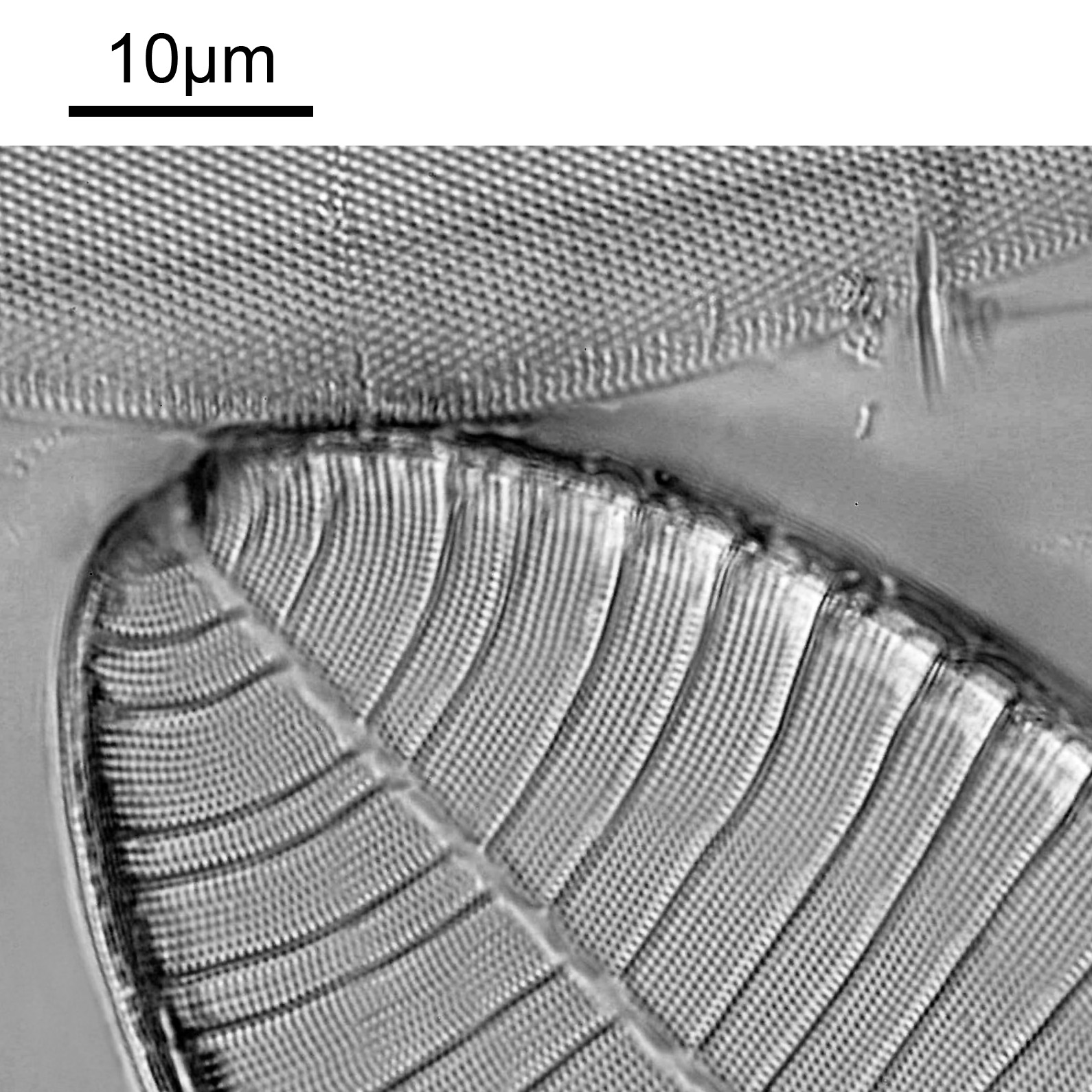
The subject will be a Surirella gemma diatom as this has some really fine structures. Here it is imaged with 313nm UV light with a 100x NA 1.2 objective.
And now an image taken using 450nm light with an NA 1.4 objective.
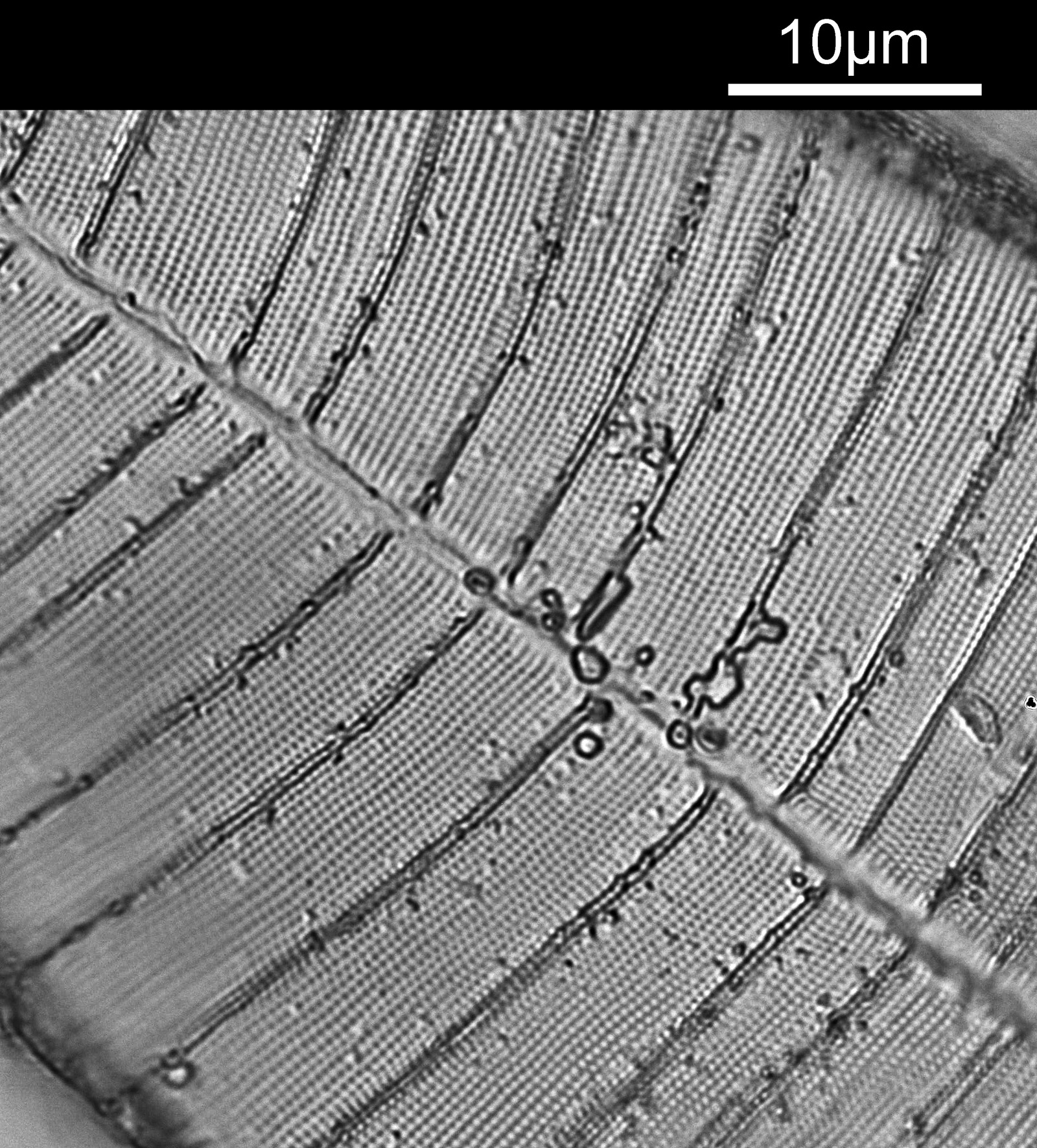
As you’ll have no doubt noticed the images have a different field of view. So a better comparison is to look at a crop from each image (both have been cropped and then resized to 1200 pixels across). First the 313nm light image.
And second a crop from the visible light image done at 450nm.
Both images seem to offer similar resolution, although the 313nm image is a little noisier. There were many differences between these images, including objective used, condenser, camera, photoeyepiece, the 450nm image was stacked while the 313nm image was not, the slides are different as are the mounts for the diatoms. As I said, ‘apples with oranges’ rather than a true ‘apples with apples’. These differences occur as optics optimized for use in the UV are not ideal for visible light, and visible light optics wont work at 313nm. However the aim with this is to compare a visible light with my best imaging conditions with UV light with a fairly basic imaging setup. The comparison between the two imaging setups is show below.
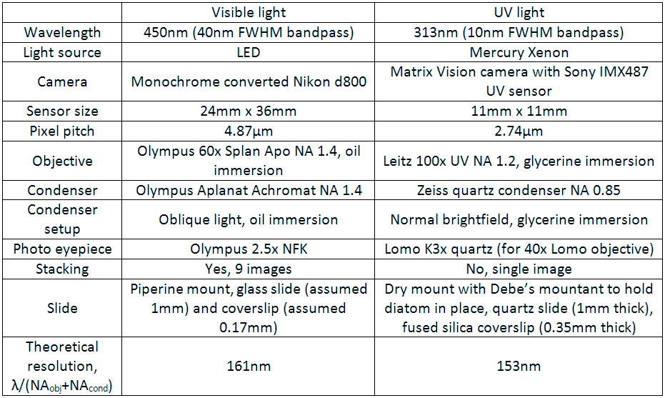
With the 450nm light I was able to use a higher NA objective and condenser compared with 313nm light. I used oblique light at 450nm, which helps resolve small features, and it was a stack of 9 images. The slide mount was also different – a nice high refractive index one of piperine, vs a dry mount in Debe’s for the UV image.
The interesting thing though is when you look at the theoretical resolution based on the wavelength of light used and the NA’s of the condenser and objective. They are very similar, less then 10% apart, with the UV objective being only slightly better. This explains why the images look pretty similar in terms of how they are resolving the small features. ‘But’, I hear you say, ‘you said that UV offers better resolution, so what is going on?’. The NA’s of the objective and condenser for the UV image were lower than the ones used for the visible light image, and this is offsetting the effect from the shorter wavelength. Visible light images can absolutely provide amazingly high resolution images of diatoms.
So, is there no point to UV imaging? Well, it’s not quite a simple as that. The NA of the condenser I used for the UV image was only 0.85. I do have a NA 1.25 one as well, I just didn’t use it as until recently I didn’t have a suitable mount for it. If I used that at 313nm, with the NA 1.2 objective, the theoretical resolution then goes down to 128nm. If I used the NA 1.2 objective and NA 1.25 condenser with 254nm light, the theoretical resolution improves again to 104nm.
Of course there are various other technique to improve visible light images, such as differential interference contrast, cross polarization and phase contrast which I haven’t looked at here. As a result modern techniques can certainly provide very very high resolutions, and the advent of these techniques was a contributing factor to the decline in the use of UV imaging for microscopy. However UV can offer high resolutions in a very simple optical setup – no need or DIC, cross polarization – and without complex image process. UV can offer these high resolutions with simple brightfield microscopy, and this is why it still has a place in imaging today.
As always, I hope you enjoyed this post, and thanks for being part of my research journey for 2022. Here’s to a better 2023, and if you’d like to know more about this or any aspect of my work I can be reached here.
EDIT 26/3/23. The slide used for the 450nm light here is labelled ‘piperine’. Piperine is an alkaloid found in black pepper, and it has always confused me as to why it would be used as a microscope slide mountant. However its use is mentioned in Microscope & Entomological Monthly, SH Meakin, The study of diatoms, 1939, Pages 209-212.