
For those who frequent my blog, you’ll have heard of the slide maker Horace Dall before. I’ve been a keen collector of his metal and metal oxide coated diatom slides for a while (and am currently writing an article on them which I hope to publish later this year). I recently came across a slide of his for sale which was a bit different though – not a diatom slide but a whole Soldier Beetle. Today I’ll share some images from this slide using both visible light and Infrared (IR).
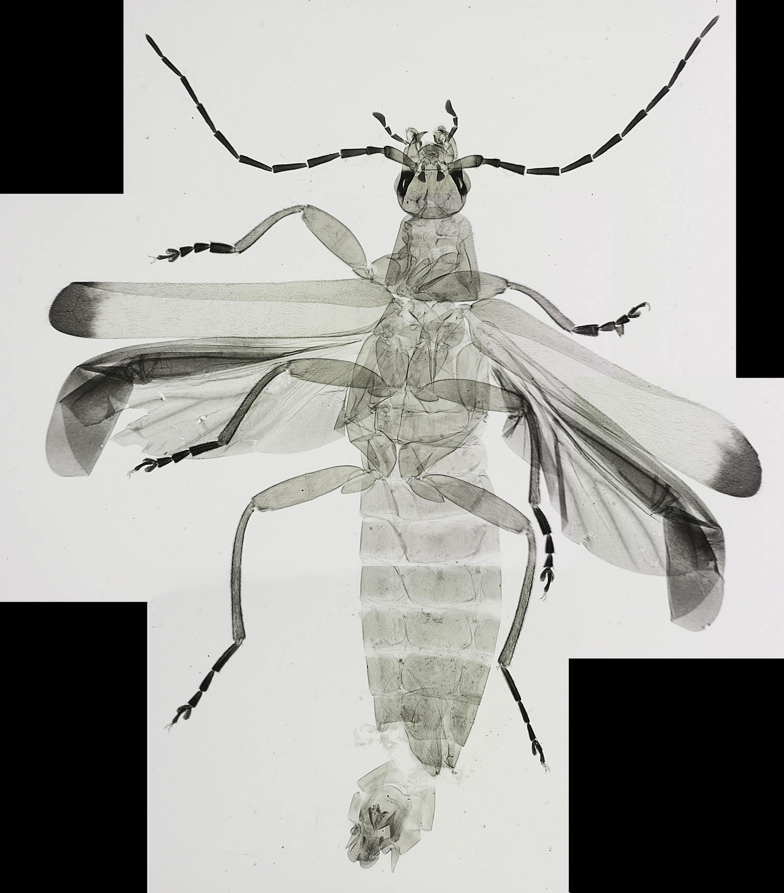
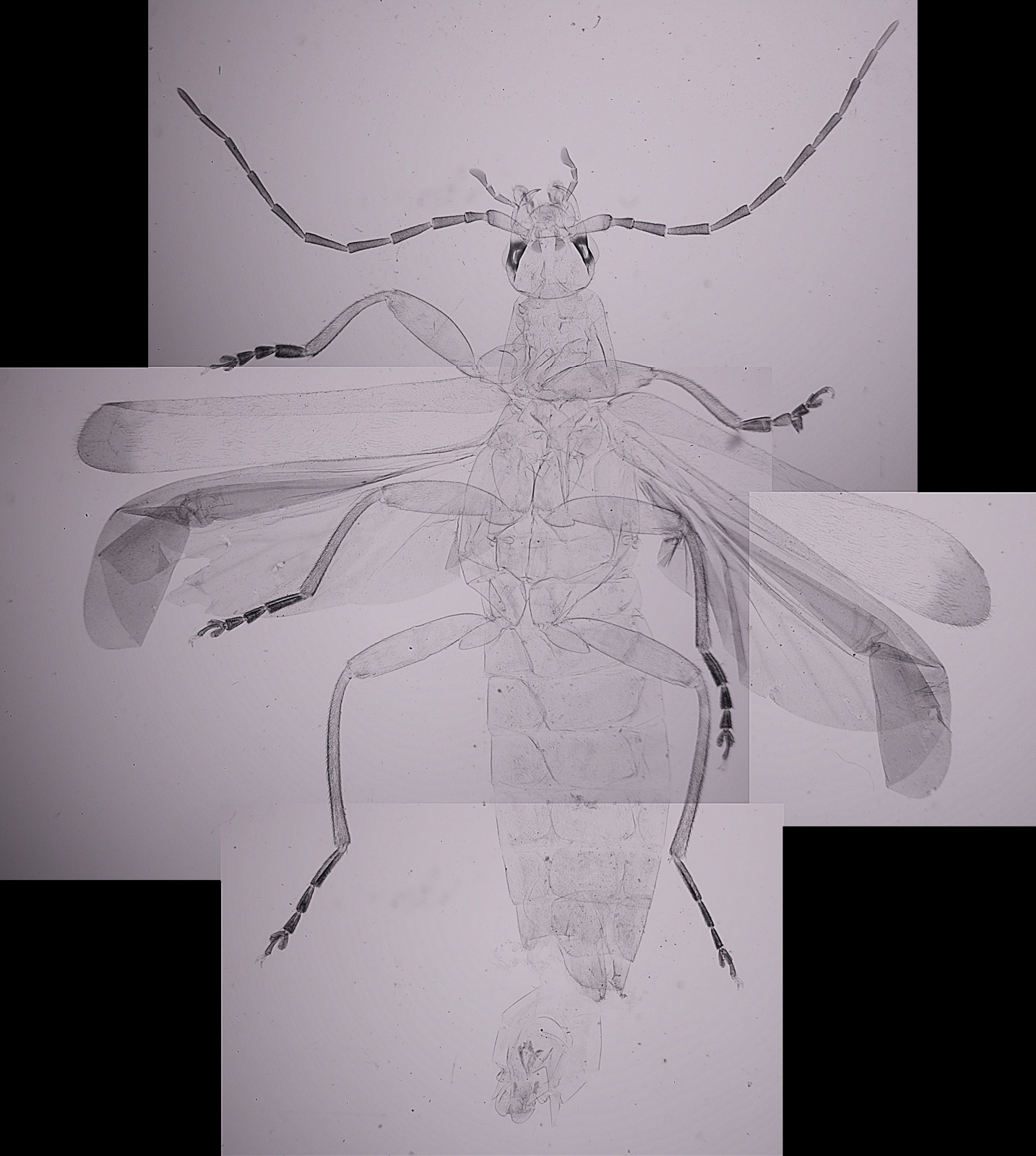
With diatoms, I normally have to go to high magnification to see them. However with this slide the problem I had was going to a low enough magnification, as the whole sample is about 20mm across. To image the whole beetle, I took 4 images using a 1x Olympus Splan Fl objective and stitched them together in Photoshop. Here’s what the sample looks like (resolution has been dropped from the original 9390×10687 to 1600×1821 for sharing).
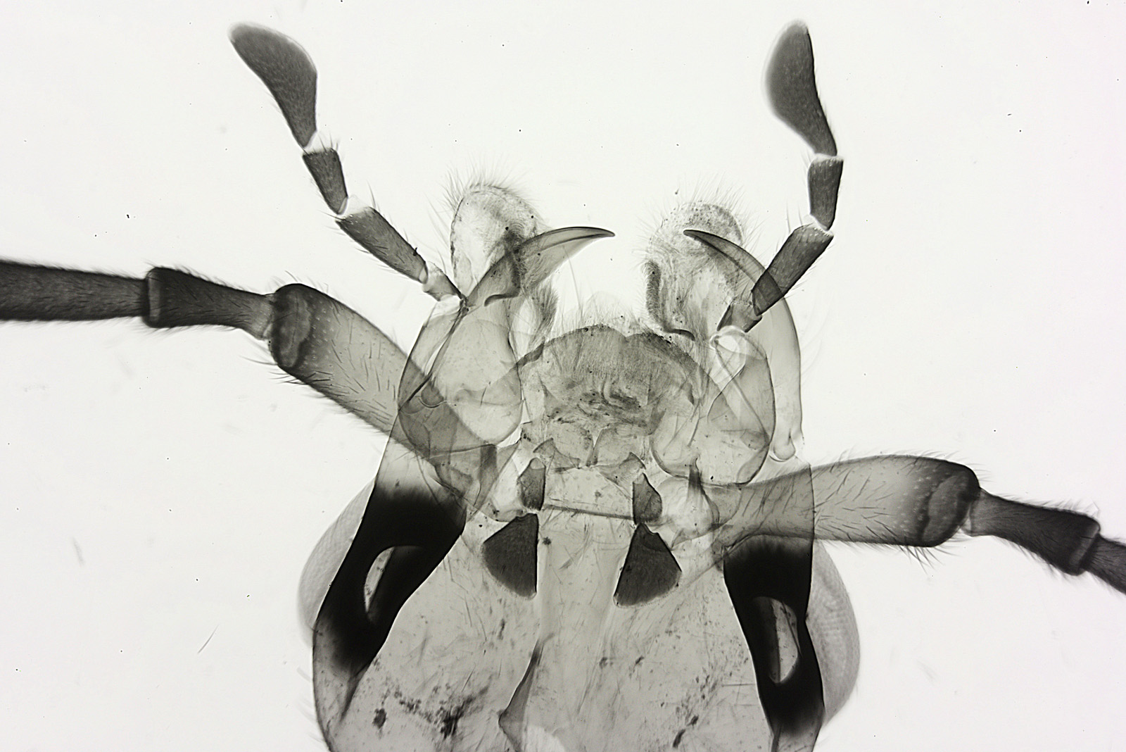
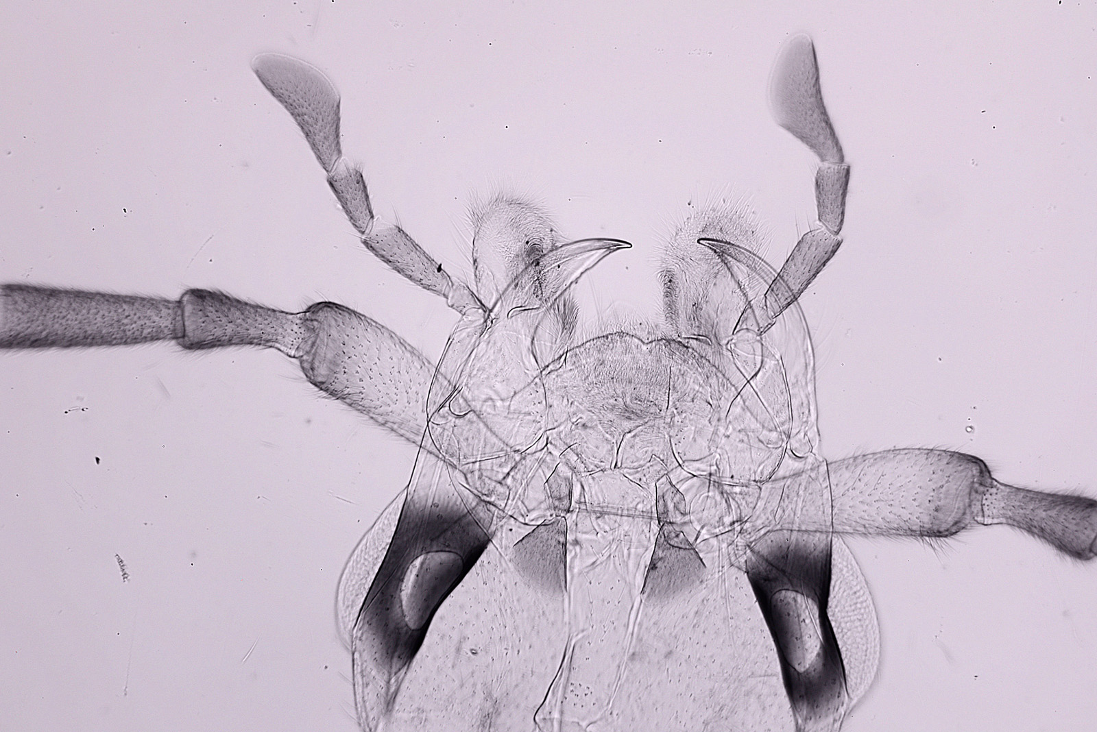
A higher magnification image of the head of the beetle using a 4x Zeiss Planapo objective.
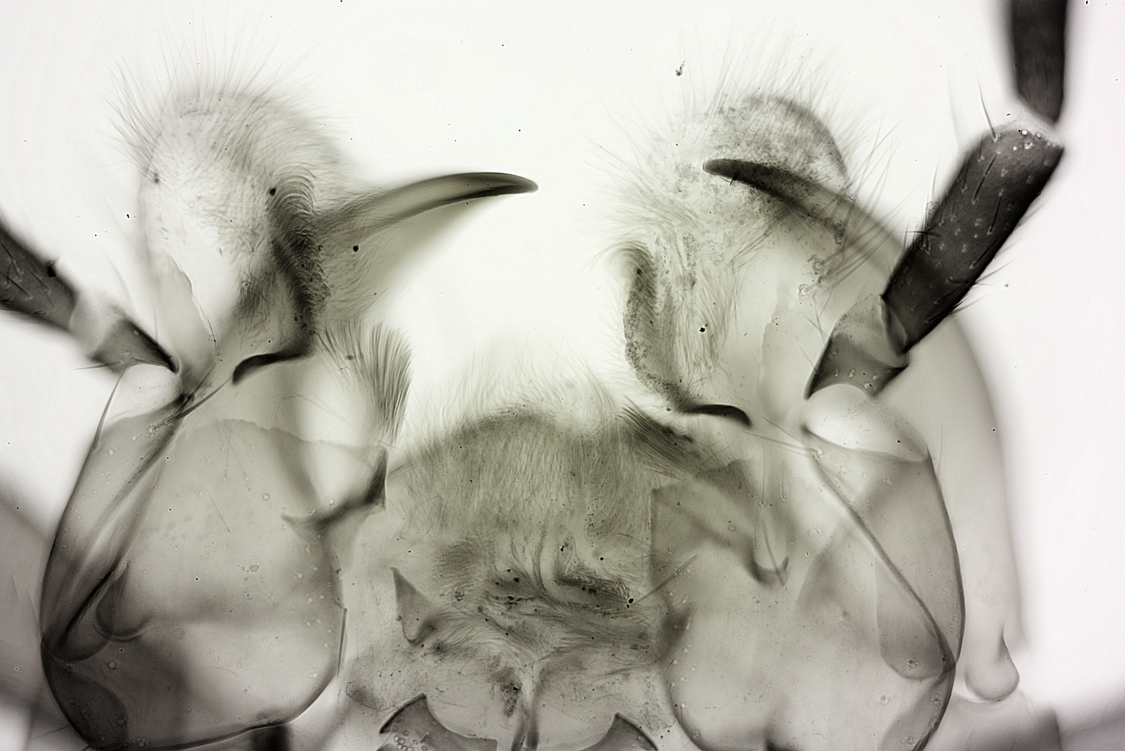
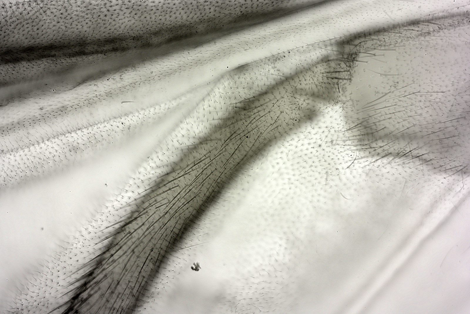
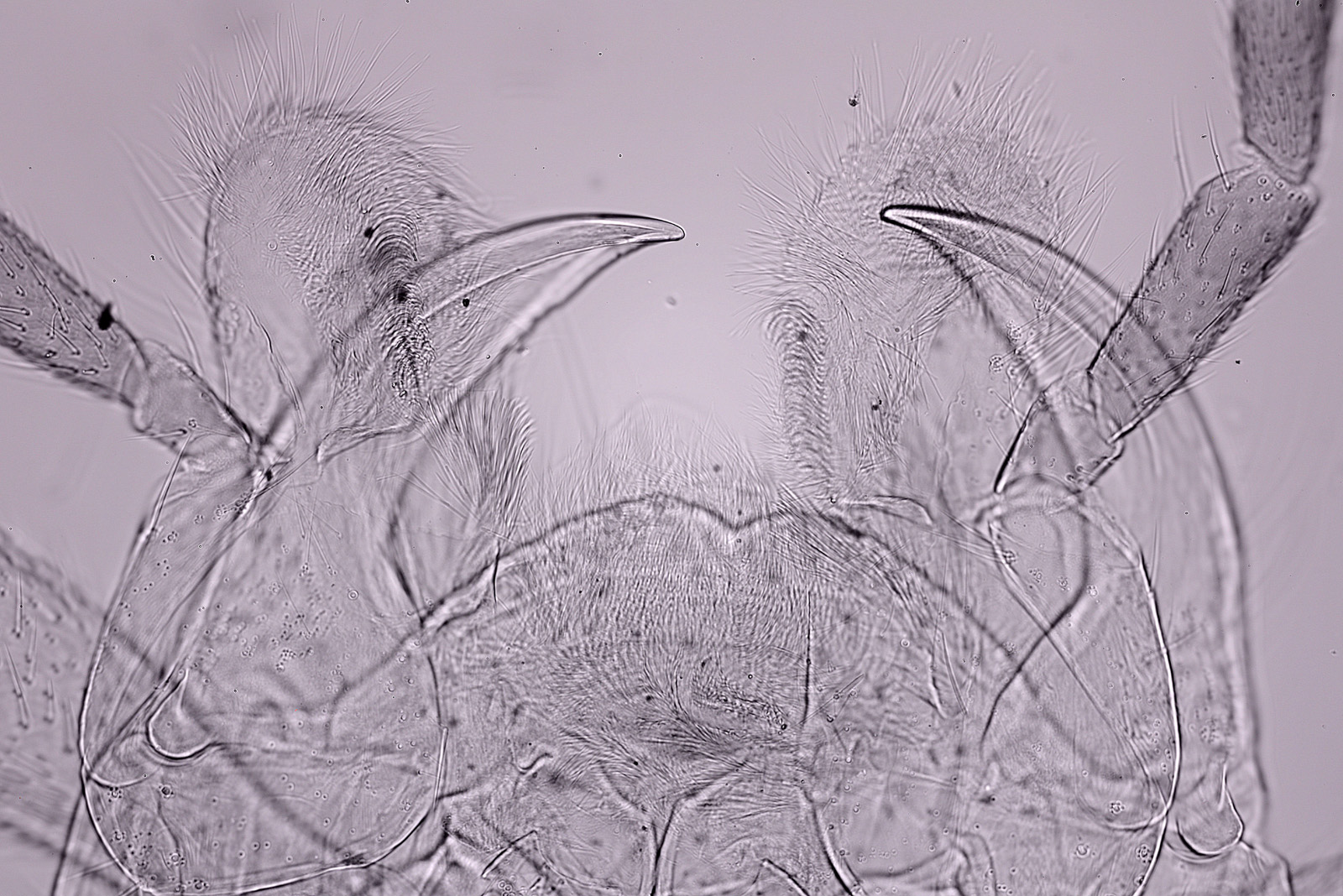
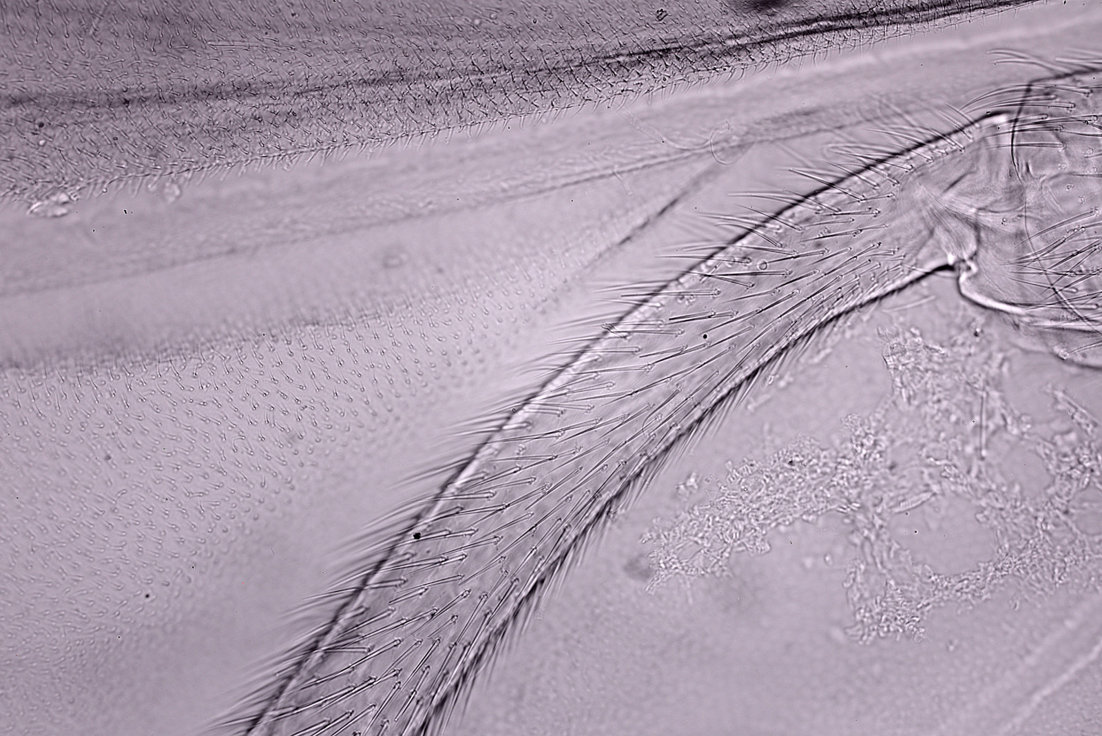
And to finish with a couple of images using a 10x Olympus UVFL objective.
While chatting with a friend on a microscopy forum he mentioned to me that IR light can be useful for imaging insect microscope slides as it makes some parts become more transparent. To try this out I swapped the LED light source for a Tungsten bulb, and placed a Heliopan 780nm long pass filter over the field lens, and then imaged the slide again.
Here’s the composite of 4 images using IR light (the IR images have a slight pink tinge in the JPEG for straight from the camera).
The IR light does indeed make parts of the insect appear more transparent, and the overall effect is to produce a lower contrast image. Next up the head using the 4x Zeiss Planapo objective.
Again, the increased transparency if obvious. Finally some images using the 10x Olympus UVFL objective.
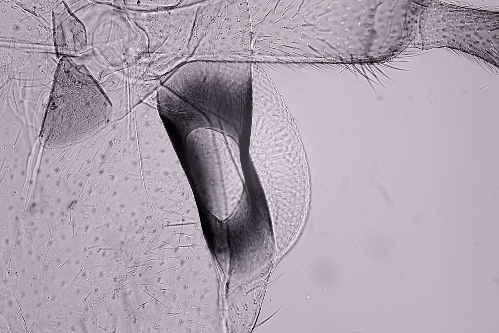
I did an additional image here of the eye with the 10x objective. The IR cuts through the darkly pigmented area and shows more of the structure of the eye. I think I need to come back to this with a higher magnification and try a stack. The 10x objective has a bit of a hotspot in the middle of the image in the IR photos. This behavior is often seen in IR photography when the lenses aren’t suitable for IR, and I presume that is the case here.
As a final set of images, I thought I would try imaging the eye region in IR with a 20x objective (a 20x Nikon Plan Apo NA 0.65). 3 different images are shown here; a single image at a given focus point (bright field), a stack of 4 images (bright field), and a single image using oblique bright field. These images were desaturated to remove the pink tinge from the IR images.
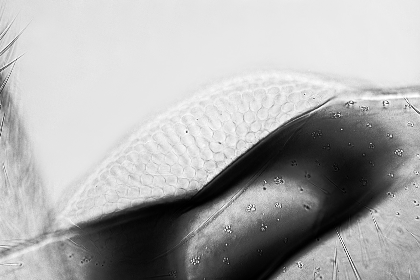
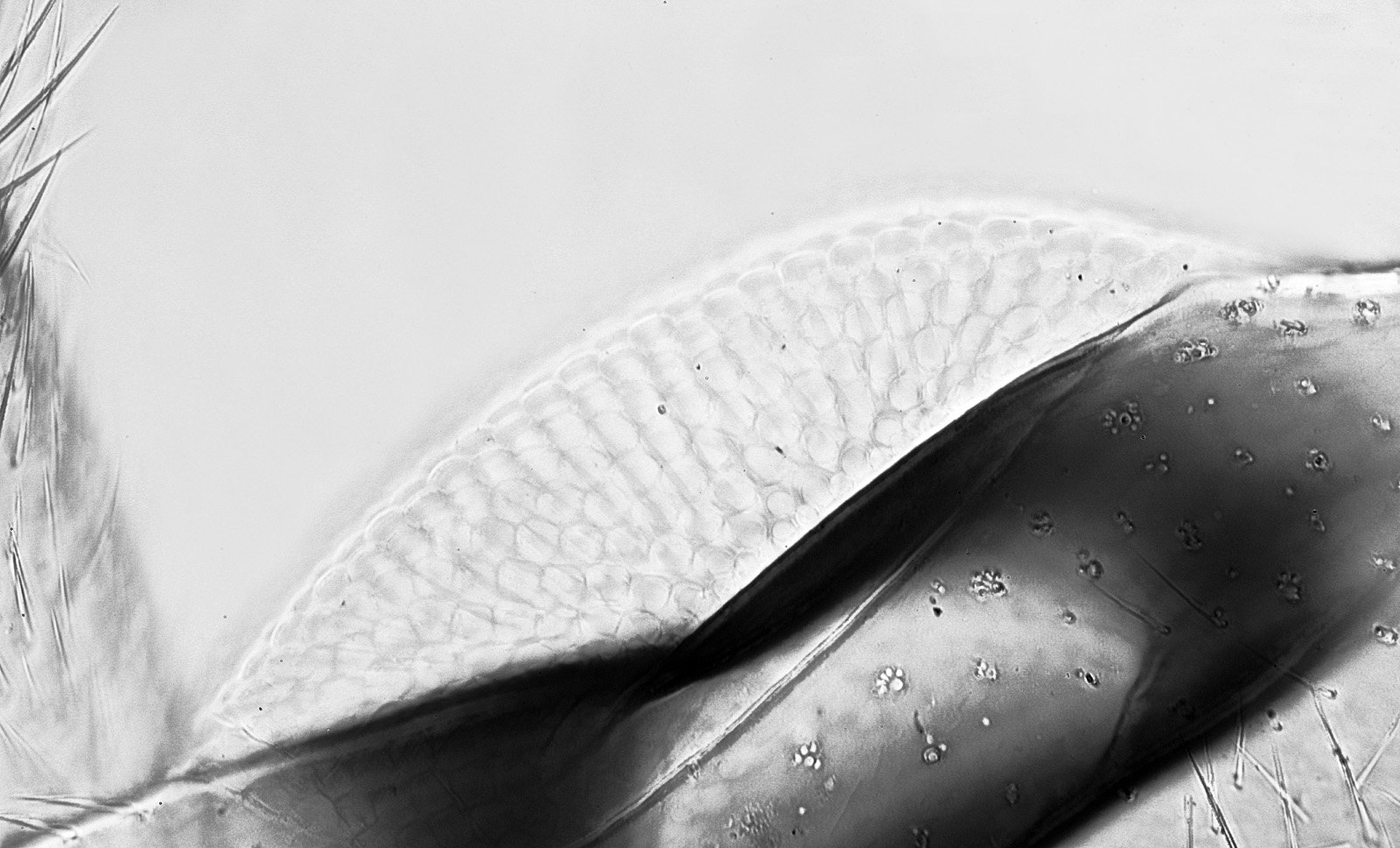
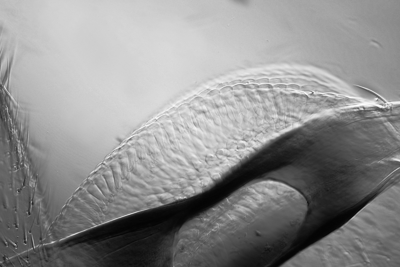
The single image using the 20x objective shows a surprising amount of detail especially in the surface of the eye. The stack of 4 images shows even more detail and some of the tubular structures of the eye lens elements. The oblique image is more of a cross section through part of the eye but does nicely highlight the structure of the individual lens elements. Interestingly this 20x Nikon Plan Apo seems to have very little hotspot in the IR so could be a good one to use again in future.
As per usual here’s an image of the slide as well.

Every now and then it is nice to have a break from imaging diatoms and look at a different sample. By moving from visible light to IR it was possible to seem through some of the dark areas of the beetle’s structure. Thanks for reading, and if you’d like to know more about my work I can be reached here.