
A while ago I shared an image from a diatom which had been sputter coated with gold to help provide increased contrast (see here). Since then I have been working on a paper about metal and metal oxide coated diatoms for microscopy which I am aiming to publish later this year. In the mean time I’ve been talking with a nice gentleman who coats diatoms with gold using sputter coating and he has sent me a few slides to examine. I’d like to share some of the images from those slides today.
For background, my modified Olympus BHB microscope was used for these, and the objective was a 60x Olympus Splan Apo NA 1.4 oil immersion one. 450nm light was used to illuminate the samples from below. Stacking was done using Zerene. Other details are shared for each image and the image sizes have been reduced for sharing here.
Please forgive me if the names of some of these are wrong, I am still learning the complexities of diatom nomenclature. First one, a Stephanopyxis diadema Ehr. from a fossil diatom strew from Dunkirk, Maryland.
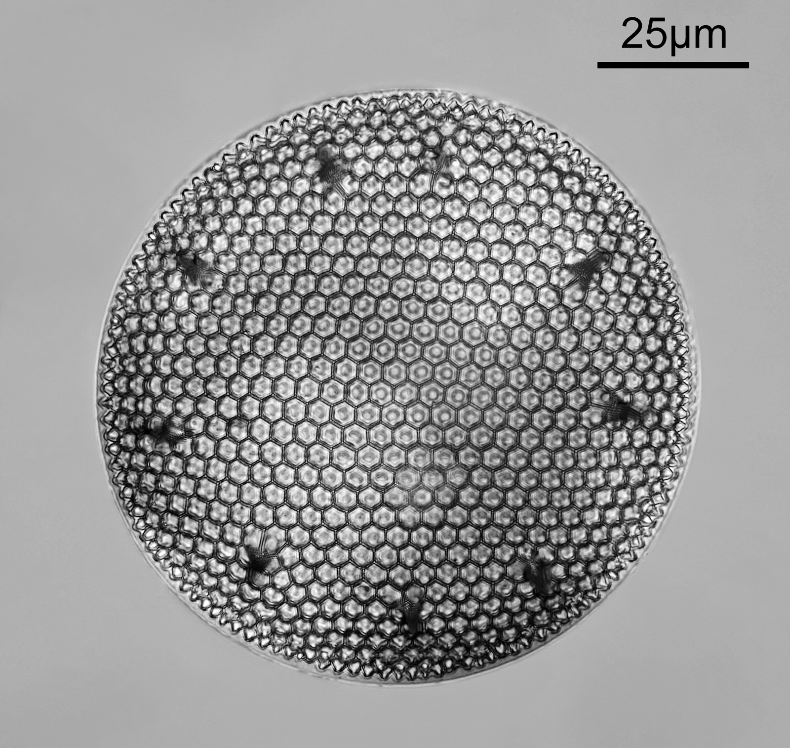
This has an amazing 3d structure, and looks to be hemispherical. There were 13 images in the stack. The tiny depth of field from the objective can be seen if you look at a video where the stage is moving.
Next is a Stauroneis (perhaps a phoenicentron) from Mud lake, Ottawa, Ontario.
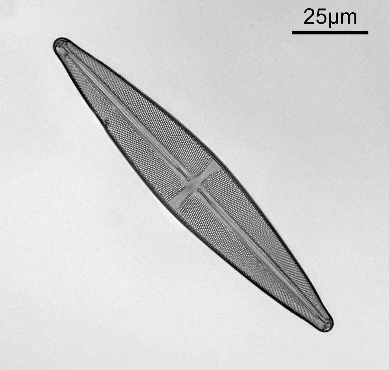
This was mounted in Pleurax and was a lovely slide to image as there was virtually no debris around it, and the diatom was almost flat. Only 3 images were used in this stack.
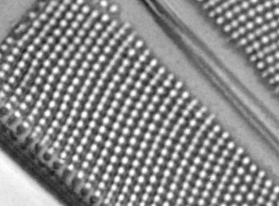
The small features don’t show up too well in the file which has been resized for sharing, but this is what they look like at original pixel resolution.
Final one is a Heliopelta of some description, again from a fossil diatom strew from Dunkirk, Maryland.

Why do this process? The gold sputter coating of the diatoms makes the silica of the structure darker and so improves their contrast, so that even simple bright field imaging (as used here) can be used successfully.
As always, I remain fascinated by these amazing structures and the worlds that can be viewed with a relatively simple light microscope. Thanks for reading, and if you’d like to know more about my work I can be reached here.