
Have you ever wondered what the inside of a microscope objective lens looks like? Why am I even asking that, of course you have, haven’t we all. All the tiny components, the little lenses. Very cool. Sometimes we get to see schematics of what they look like in cross section in manufacturers brochures or in other publications, but usually that is as close as we get to actually seeing what is going on in there.
I recently saw an objective for sale which had been sectioned from end to end by Spectrographic Ltd in the UK, so put in a bid and was lucky enough to buy it. The objective was an Olympus HI M100 1.30 which was unfortunately broken (no working lenses were sacrificed in the making of this section). Looking from the side it looks ok, a bit beaten up perhaps, but ok;

However turn it around and it looks very different.
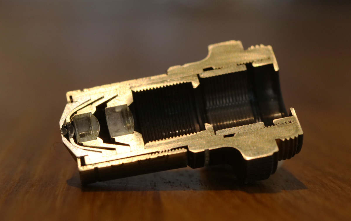
Keep in mind that this objective lens is only about 25mm long. By sectioning it the amazing engineering that it takes to make one of these is revealed. The lens elements themselves are remarkable, as can be seen when we zoom in on them.
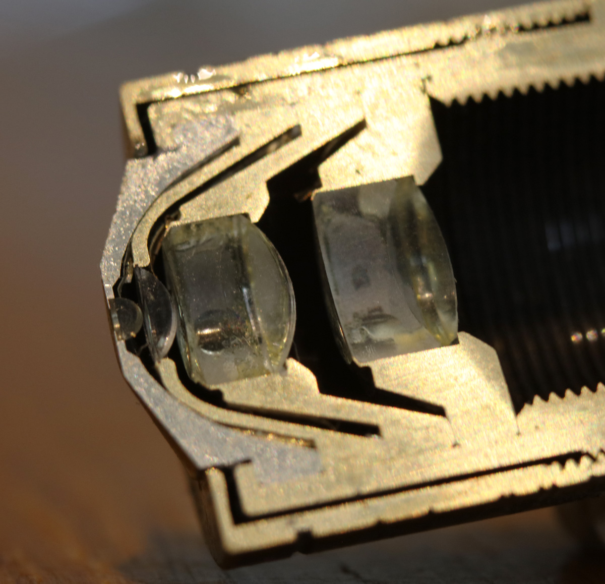
Going from left to right it looks like there are two singlets, and then two cemented doublets. Keep in mind that these are only a few mm across.
The images were captured using a Hoya Super EL 60mm enlarger lens as a macro lens as discussed here.
I’m very impressed by the quality of the section from Spectrographic Ltd, especially considering it is such a small item. This is something that is handy to have when explaining about microscopy – being able to show someone the inside of an objective is a step up from just looking at a sketch on a screen. Now then, where is that broken Beck reflecting objective I got a while back…..
Thanks for looking and if you’d like to know more about this or my other work you can reach me here. And please remember that the images in my site are copyrighted to JMC Scientific Consulting Ltd. If you’d like to share them, please ask first.