Category Archives: General

Microscopy – Mystery of the Leitz 50x Pv glycerine immersion lens solved

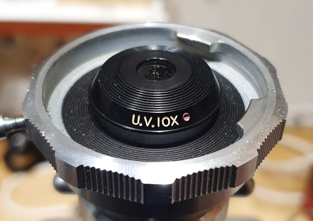
This is something which goes back to my early days of exploring microscopy in 2020. I bought a slightly mysterious lens, a 50x Leitz Pv NA 1.0 glycerine immersion objective, which has designed to be used with a 0.18mm thick quartz coverslip. I originally wrote about this here. At the time when I tested it I was slightly surprised to find that despite being designed to be used with glycerine as the immersion fluid and for use with a thin quartz coverslip, the UV transmission was not like a Zeiss Ultrafluar. Although offering some UV transmission especially at 365nm, it dropped at the shorter wavelengths and was blocking the light by about 320nm. At the time I didn’t expect this, as I had thought it would transmit further into the UV, however the objective had some unusual markings on it, and I wondered if this was a prototype, mock up or master copy for the factory perhaps just fitted with glass elements. Since then I have always kept a look out for another copy of the lens to test for transmission and compare with my original copy. However this is a rare objective, and it was nearly 4 years before I saw another one for sale. Today’s post shares the results of my transmission testing of this new copy, and discusses the implications of my findings.

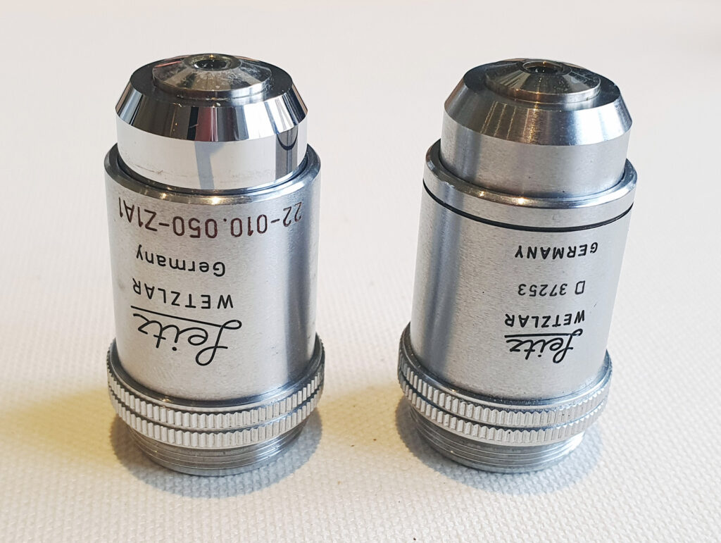
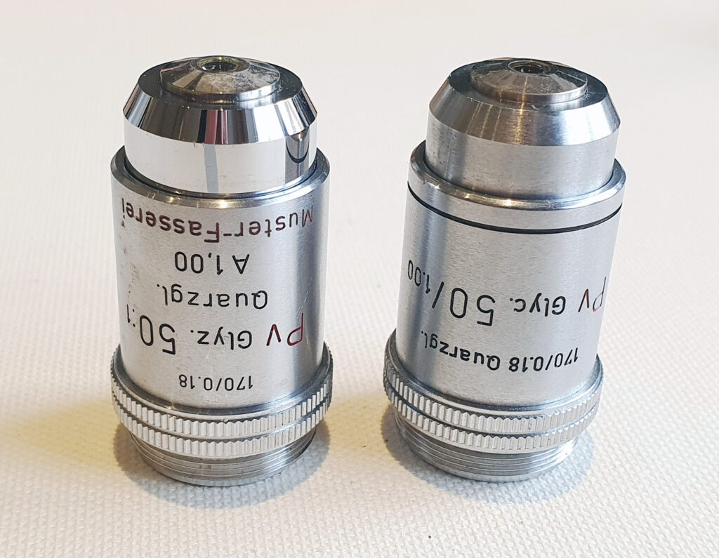
Here are the two copies of the objective. My original one with ‘Muster-fasserei’ written on the side on the left, and the new copy on the right.

Two 50x Leitz Pv NA 1.0 objectives

And the back side of the objectives.

Two 50x Leitz Pv NA 1.0 objectives, back side view

There are some differences in style and labelling of the two objectives, but overall they should be the same – 50x magnification, phase contrast (Pv), NA 1.0, glycerine immersion and for use with a 0.18mm thick quartz coverslip on a 170mm tube length microscope. Except, according to the original Leitz literature, they were actually meant for a microscope, but a ‘microspectrograph’. However they do work as normal microscope objectives with a phase contrast ring in them.

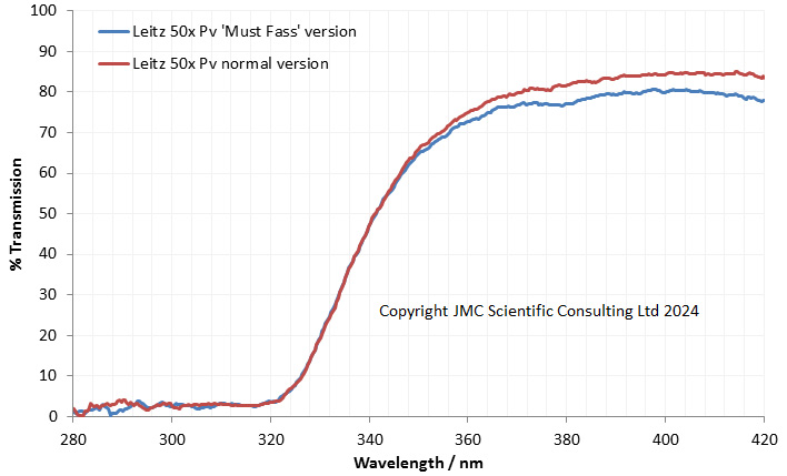
So, the six million dollar question, how does the UV transmission of the two version compare? To check this I measured both of them using my Ocean Insight FX spectrometer and got the following graphs.

Transmission through the two copies of the 50x Leitz Pv objectives

What does the graph above show? There are the two copies of the objective. The transmission through the original one with ‘Muster-Fasserei’ in blue, and the new copy in red. The transmission spectra for the 2 objectives are almost identical and certainly close enough for me to be happy that there are no major differences between them in terms of construction. Both have good transmission at 365nm, and then dropping in transmission below that, with them being essentially opaque by 320nm. Below 320nm we are down in the noise and although it says about 3% transmission this is just a quirk of the measurement process.

Where does this leave us? My original ‘Muster-Fasserei’ copy has the same transmission as the other copy, so optically I am happy that this is the same as the production copies. They have good UV transmission above about 350nm, but poor transmission below that, so they are not true UV lenses like Zeiss Ultrafluars or the Leitz UV objectives, but then they do not claim to be. Glycerine immersion normally would indicate UV work, and the 0.18mm thick quartz coverslip would also back that up. Now though I come back to the entry in the Leitz catalogue which described the objective for being for a ‘microspectrograph’. I suspect the use of glycerine as an immersion fluid was to reduce fluorescence as it is low fluorescence fluid. Also the quartz coverslip will be lower fluorescence than a glass one (I’ve looked at fluorescence of glass and quartz/fused silica here). Minimizing fluorescence of components in the optical train will be very important for a spectrograph which will be measuring light intensity. These were therefore probably designed to be used in a setup where low fluorescence was important, so perhaps for use with light with wavelengths from 350nm and up.

Answering questions about older microscope equipment can be a real challenge. Documents do not always exist online, and personal knowledge of folks who might have used or worked on older equipment is unfortunately disappearing rather too rapidly. This has taken a few years to get an answer but I feel I know more about them now than I did before, and as a scientist that is very important to me. As always, thanks for reading and if you’d like to know more about my work, I can be reached here.

Microscopy – A diatom wish list

Firstly, apologies for not posting a lot recently. My wife and I had a holiday in Tasmania in January, and I am still going through the photos from that (it’ll be done by the end of the month, fingers crossed). I have done a bit microscopy in my down time though, and it got me wondering, what would be my wish list for diatoms to image? I’m sharing some thoughts on this today, some of my wish list I have and others that I have yet to image. These images were done using my modified Olympus BHB microscope, and have been reduced in resolution for sharing here (the original are much higher resolution).

EDIT, 18th March 2024. I have managed to find an example of one of the diatoms am looking for – Truania archangelskiana – on a strew slide I had, so have included an image of that now.

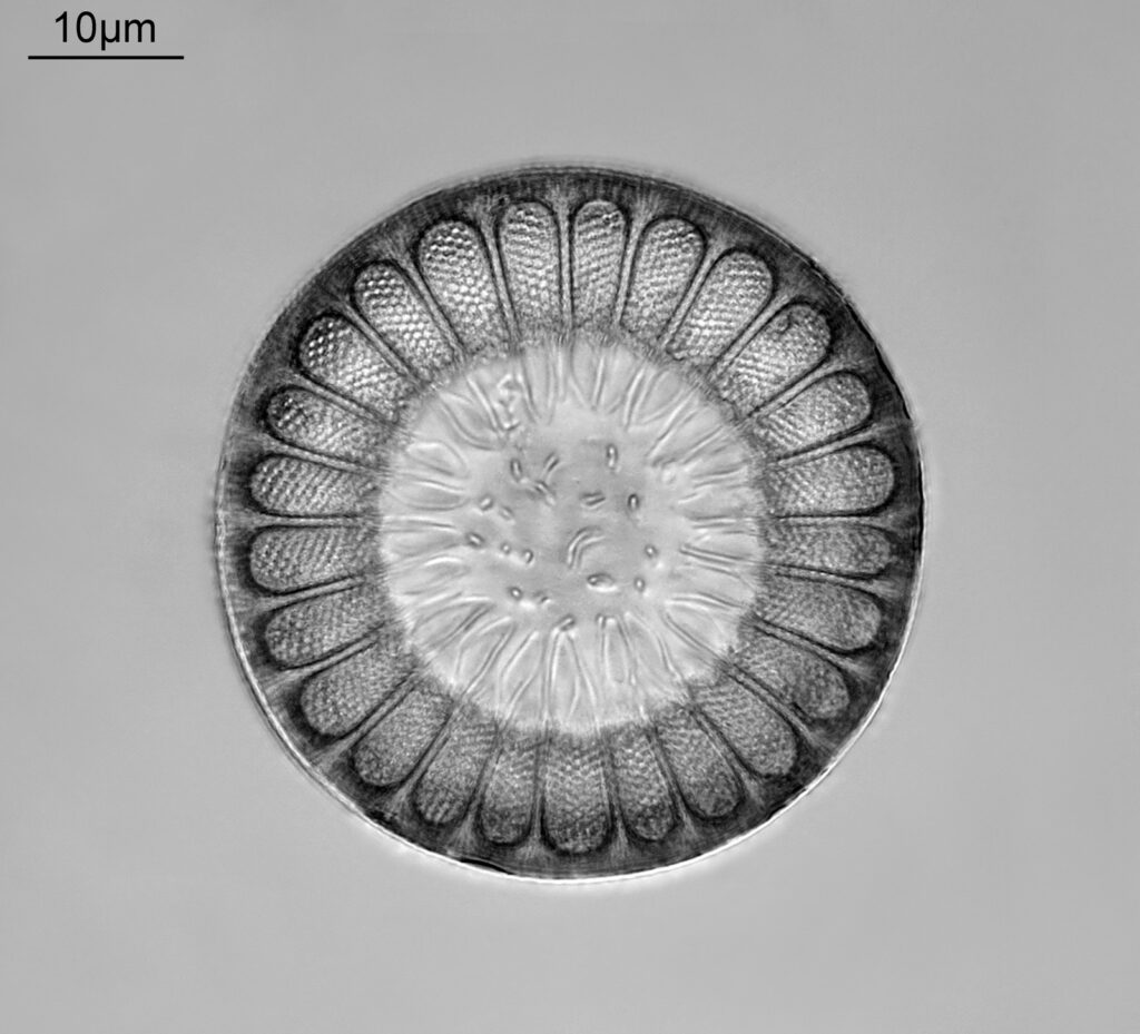
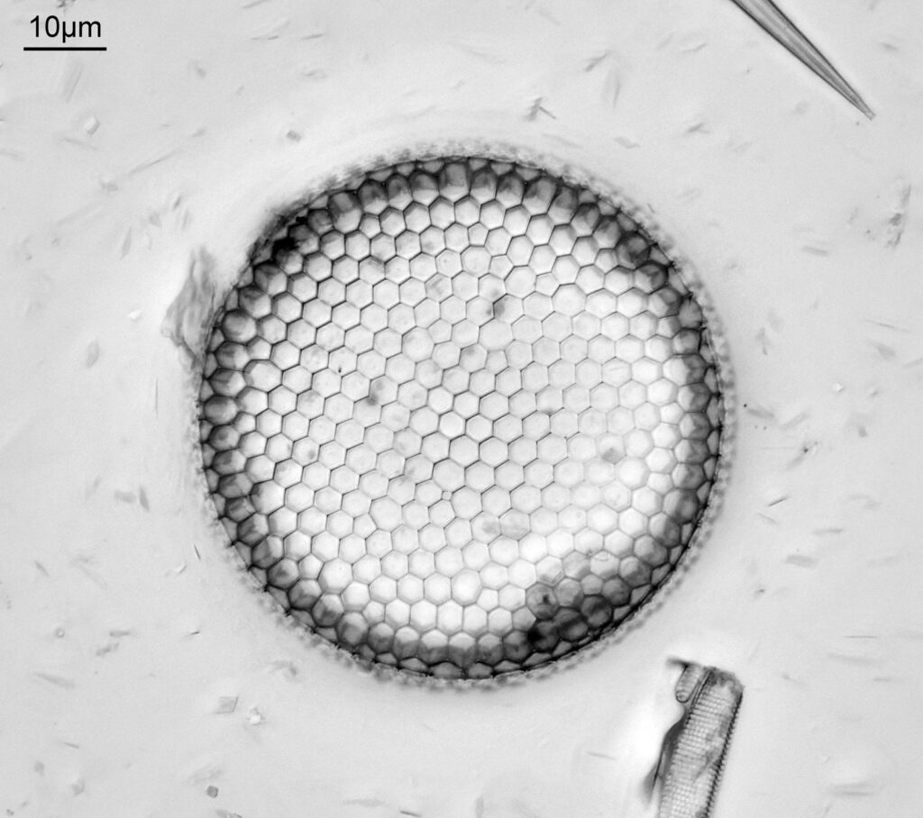
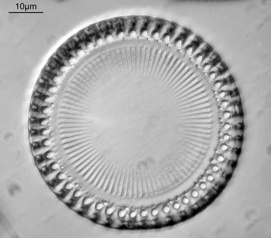
I’m going to start with ones on the wish list that I have managed to find. Firstly one of the structurally prettiest diatoms I have imaged so far, Cerataulus subangulatus from the Oamaru deposit.

Cerataulus subangulatus

This was a slide made by Emiliano Bellotti and has a single example on slide. A beautiful structure as well as being a challenge to image.

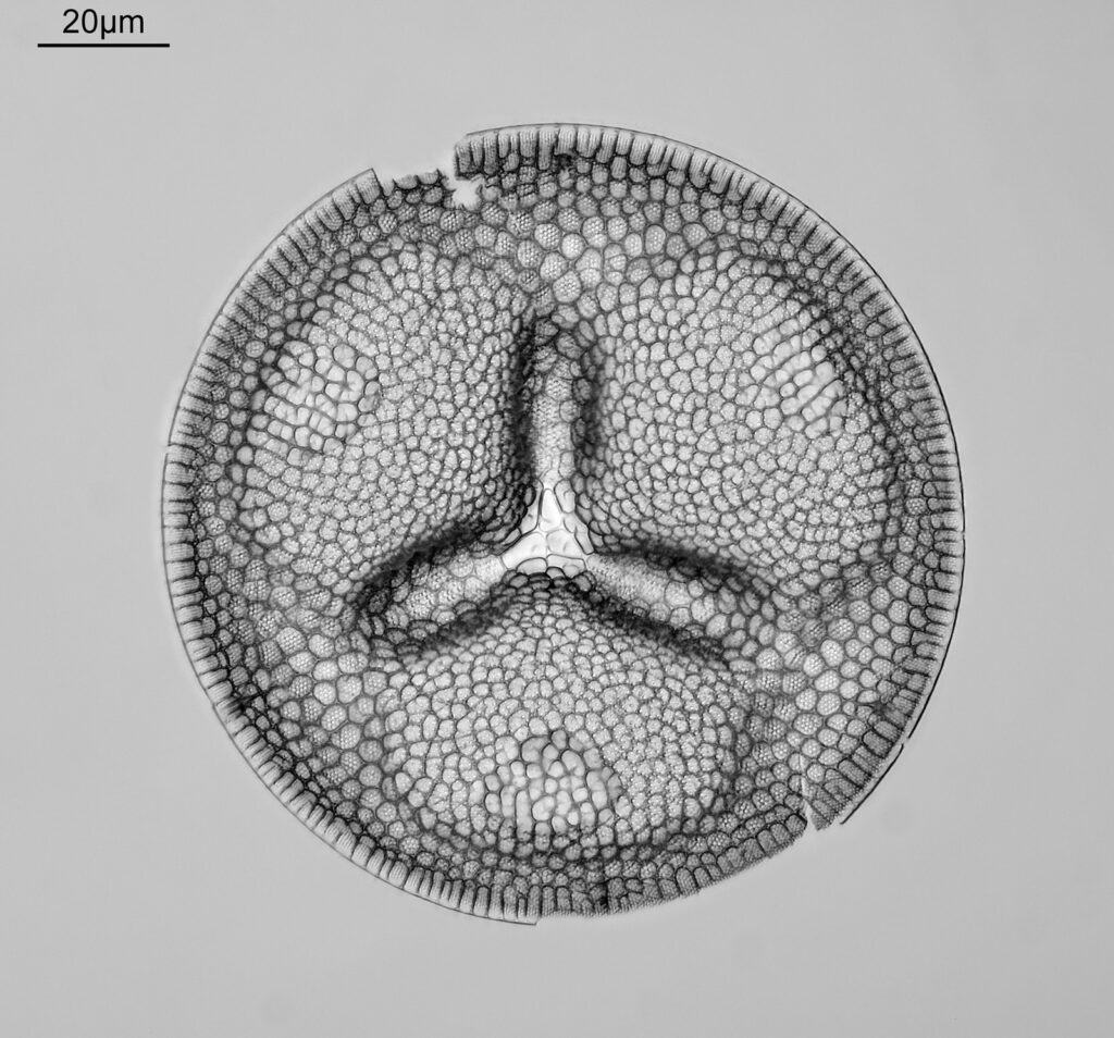
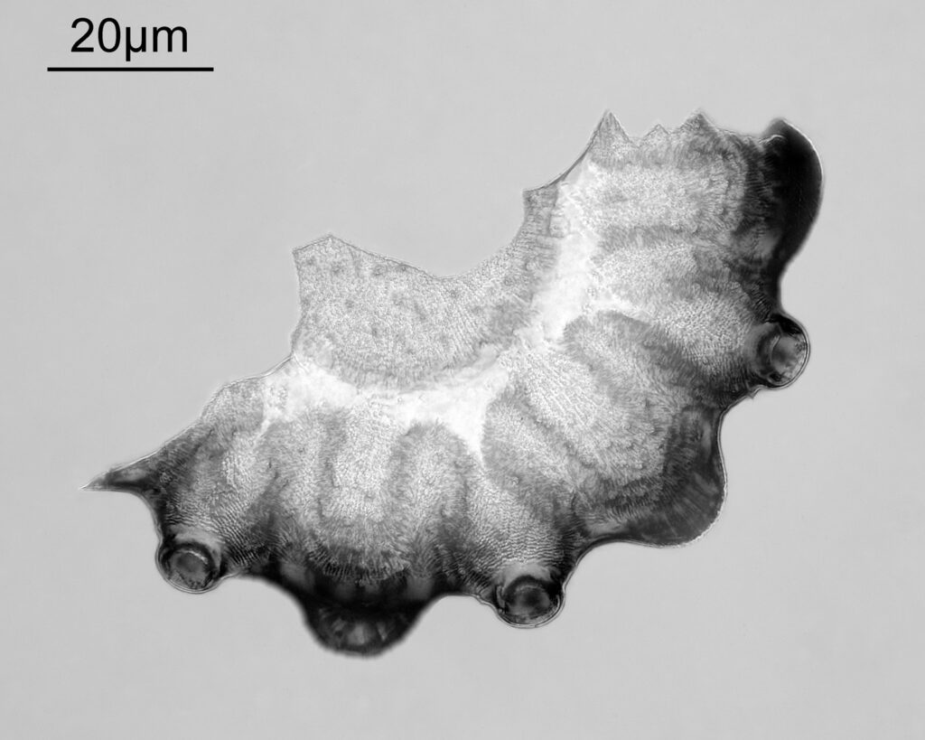
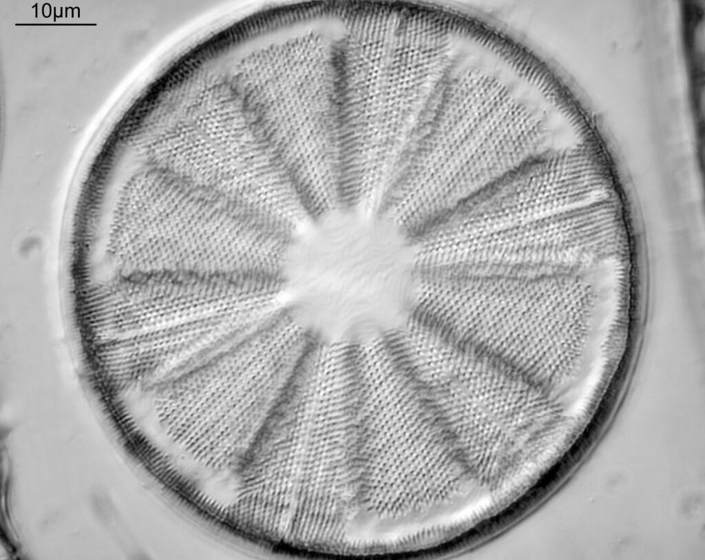
Next we have Brightwellia Coronata, again from Oamaru.

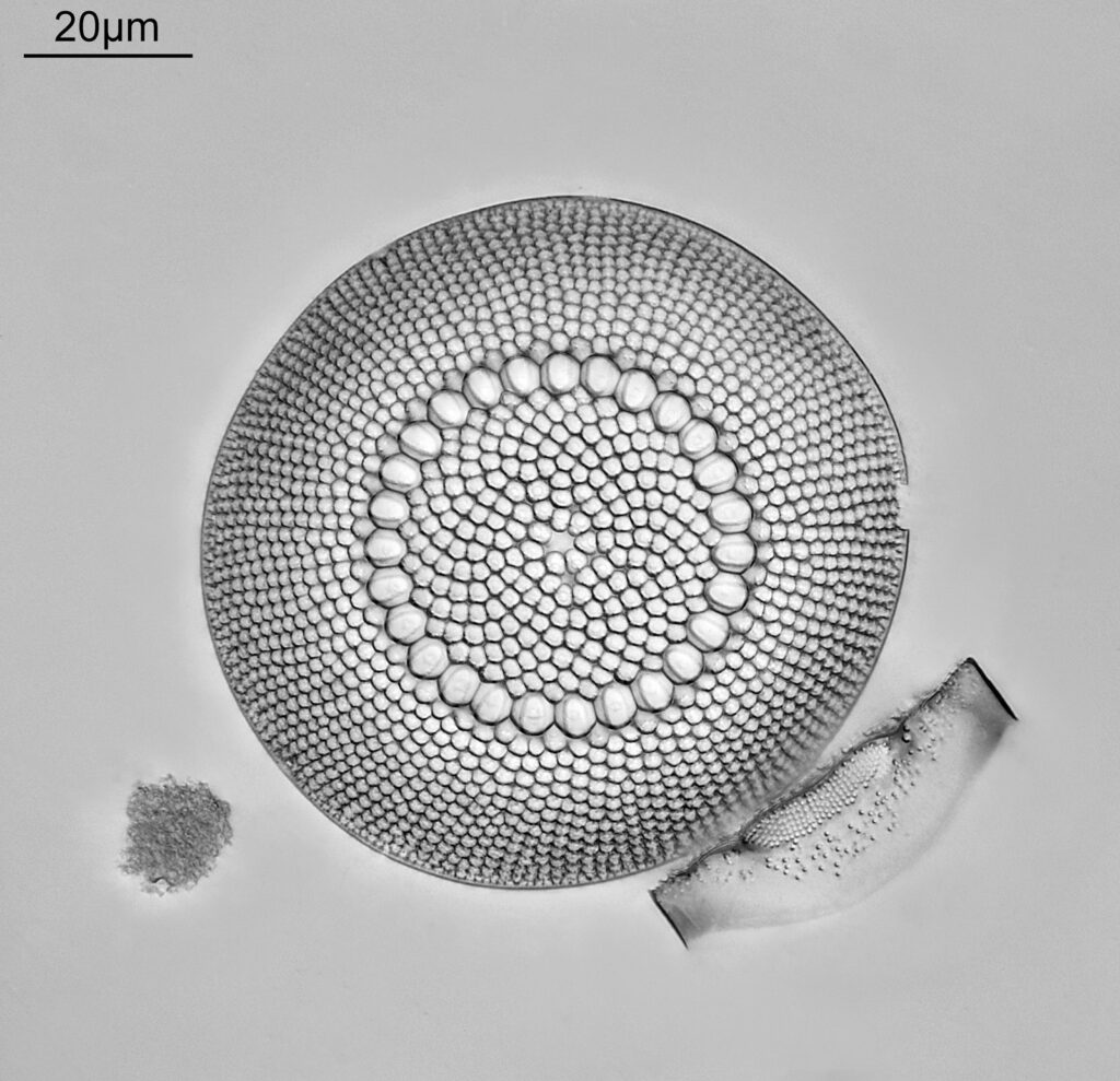
Brightwellia coronata

This was on a strew slide from Allans Farm, Oamaru by Bernard Hartley, and is the most complete one I have found in the slides I have. It seems to be very prone to breakage, and finding a complete one is a ‘challenge’, although many Oamaru strew slides will contain fragments of them.

Next is Hydrosilicon mitra.

Hydrosilicon mitra

The slide maker here is JA Long, and the location is given as New Hebrides. It’s quite a rare diatom and extremely fragile. This one is the best I have with only a few broken ribs.

Next is Monopsia mammosa, another one from Oamaru.

Monopsia mammosa

Another slide by JA Long, and I do have second example of one which is more broken up. This has a couple of cracks, but that is about it.

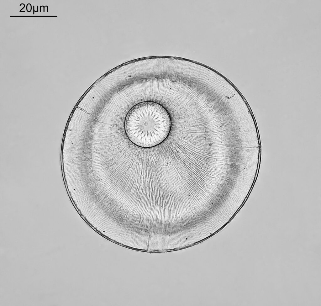
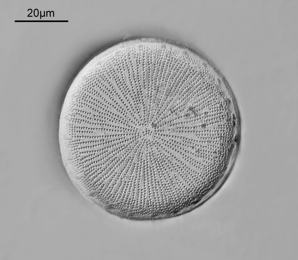
This one I only imaged a few days ago – Actinoptychus affinis from Java.

Actinoptychus affinis

This was a slide by Samuel H Meakin, and is almost complete apart from a bit of the edge missing. I love the patterning on this one.

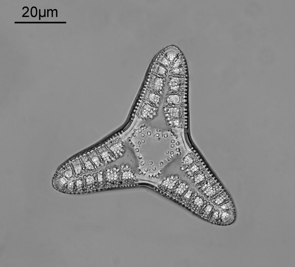
Now for Triceratium nitescens on a slide by the maker RI Firth.

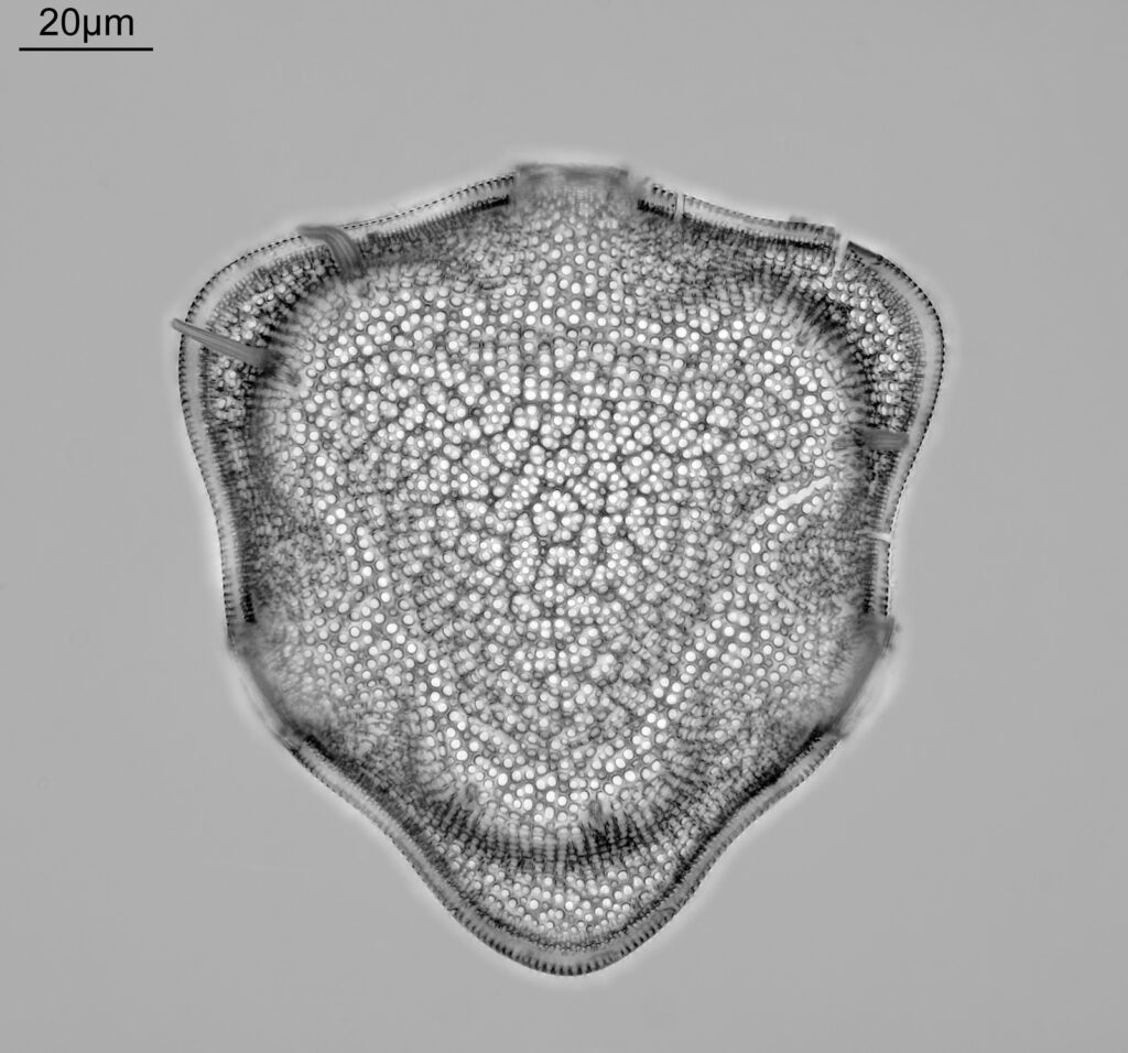
Triceratium nitescens

A beautifully shaped diatom from Barbados and one I was really happy to be able to image (the slide says ‘very rare’ and I believe that is true).

Now we get into ones that I am still actively looking for examples of, because I either only have a fragment of it, the one I have is not ideal for some reason, or ones which are so unusual I have only seen pictures of. To start with Charcotia decrescens from Antarctica.

Charcotia decrescens

This one is on a strew slide by Klaus Kemp, and is actually pretty good. I’m being picky here, it was at quite an angle in the strew, and made imaging difficult, so is one I keep an eye out for. I think it is also known as Chacotellia decrescens, and Actinocyclus actinochilus.

The next is one I only have a fragment of – Biddulphia pedalis from Oamaru.

Biddulphia pedalis

Another slide by Samuel H Meakin, I have seen fragments of it, but so far have not tracked down a whole one and it seems to be quite a rare diatom. Also known as Grovea pedalis. Lovely details on it though. The image above was done using 365nm light and the short wavelength makes all the small features really pop.

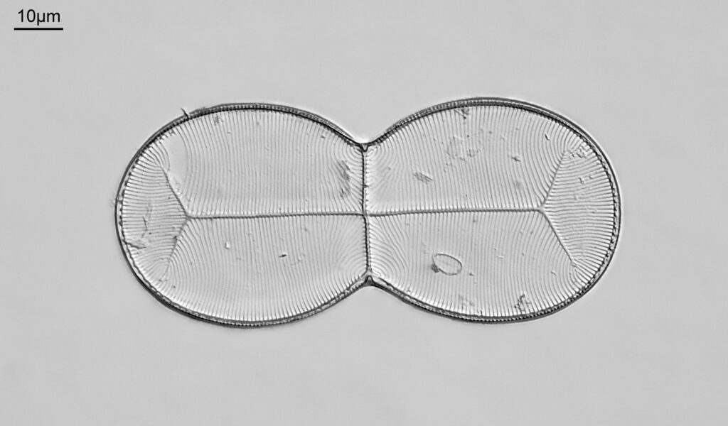
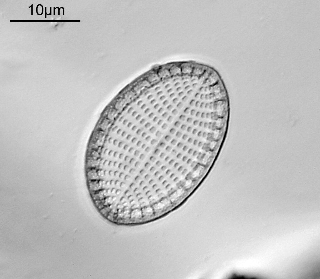
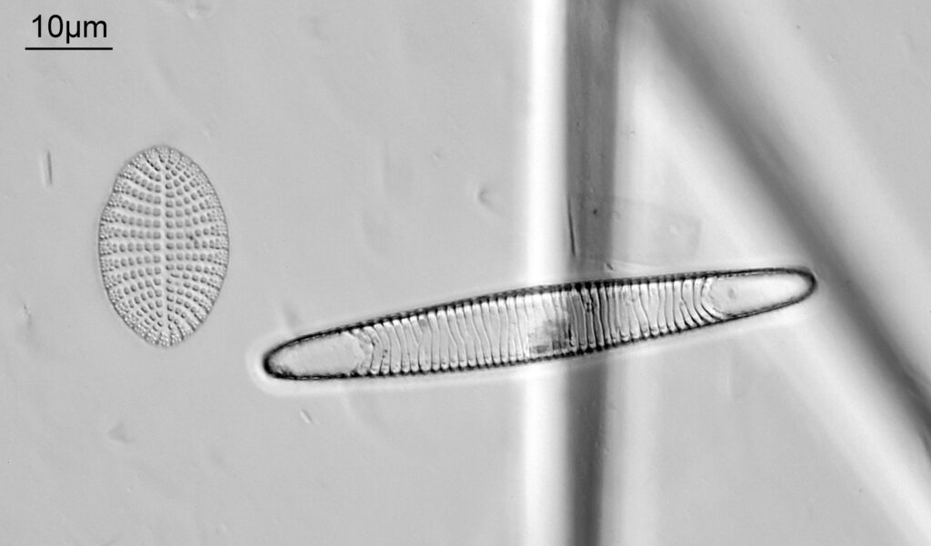
The last one I do not have a individual sample of, but I have managed to find it on a strew slide by Bernard Hartley. It is Truania archangelskiana, and is shown below.

Truania archangelskiana

Reported locations for this one include Singiliewsky and Inza, Russia and Eidsbotn Fjord, Devon Island, Nunavut, Canada (seen here). An image of one can also be seen on the Photomacrography forum, here. A striking looking diatom and well worth looking out for for imaging.

I find imaging diatoms fascinating and rewarding, and never cease to be amazed by the varying forms. There are loads I could have listed here, but this gives a nice cross section of some of the more unusual ones. I hope you enjoy these images, and if you have any slides of these you would be open to finding a new home for, I can be reached here.

UVA and UVB Video microscopy of titanium dioxide (TiO2) particles in sunscreen films

Firstly, a belated Happy New Year to everyone. I wish you all the best for 2024. I start the year with some microscopy work, but something a little different to the diatoms I regularly share. My day job is in the field of dermatology, and the initial reason I built my UV microscope was to be able to image sunscreen (SPF) products. Specifically to be able to image UVA and UVB sun filters separately and see where they are in the cream. While most of my imaging is done using photographs I do occasionally do video work. This post shares some really funky videos looking at titanium dioxide (TiO2) particles in a partially dried sunscreen film, with the videos being recorded both using UVA – 365nm light – and UVB – 313nm light.

The product contained titanium dioxide (TiO2) particles as the UV blocking ingredient. The slide was prepared and the product allowed to dry slightly before imaging. Imaging was done using my modified Olympus BHB microscope with a 100x Zeiss Ultrafluar NA 0.85 objective lens, and using two different UV wavelengths – 365nm (UVA) and 313nm (UVB). This is not fluorescence imaging, this is direct imaging of the UV at the different wavelengths.

Two videos to share. First, captured using UVA light.

UVA video of partially dried sunscreen film

What are we looking at here? The video shows a partially dried down film of a sunscreen cream. There are regions where there are still droplets of oil from the topical emulsion before the droplets have chance to coalesce. Inside the oil droplets there are particles which are moving – these are particles of TiO2. Some of these particles look white and some look black in UVA, i.e. some are absorbing the UVA and go black, and some are reflecting or scattering the UVA, and look white. Whether they absorb or scatter a given wavelength depends on their particle size. However the really cool thing is that they are moving and moving quite a lot. What’s causing this? I’m shining UV on the slide with the product film and some of that is being absorbed by the TiO2. As a result it will warm up and start to move – Brownian motion – bouncing into other particles and transferring energy causing them to bounce around. As a result UV energy is absorbed resulting in particle motion.

With my microscope I can image other UV wavelengths. By swapping filters and imaging at 313nm I got the following video.

UVB video of partially dried sunscreen film

Same region of the product film, but now we have a video of how the sample looks using UVB light of 313nm. The video is much grainier as the camera is less sensitive at that wavelength, so the ISO needed increasing to be able to capture video footage. The particles look different now – most look black showing that most are now absorbing the 313nm wavelength light rather than scattering it. This is to be expected for TiO2 particles as the absorption of light is wavelength dependent. The motion is still present. The particles are typically sub-micron in size, but are probably aggregations on individual TiO2 particles as the smallest ones would be too small for me to resolve on my microscope.

Before I wrap up, I want to revisit the second video. Excuse me while I geek out for a moment. This was imaged using 313nm light and through a high magnification microscope objective. The camera was a monochrome converted Nikon d800 prepared by MaxMax in the US. Even though monochrome conversion improves sensitivity at 313nm, I was amazed to be able to capture live video of the behavior of this sample and how it looked in the UVB region.

It’s funny, when I first looked at these samples the movement I observed reminded me of cells moving around. However as I mentioned above, this is about energy transfer and heating of inorganic particles. The motion, while cool to see, is actually a problem for capturing images. Often my exposure times for capturing UVB microscope photos are of the order of half a second or more. Movement of the particles causes blur and reduces the resolution achievable. As always it is important to understand the techniques you are using when trying to understand what it is they are telling you.

I’d like to thank DSM-Firmenich AG for supplying the sunscreen sample to image, for funding some of my UV microscopy research and for giving me permission to share these videos. As always, thanks for reading, and if you’d like to know more about my work I can be reached here.

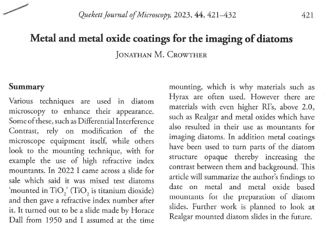
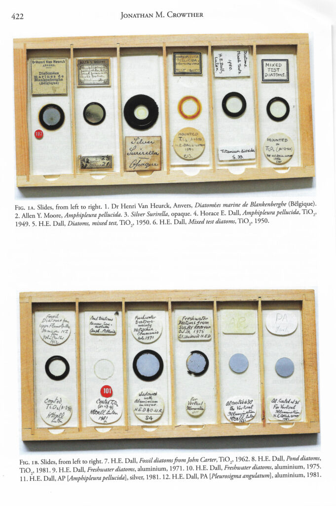
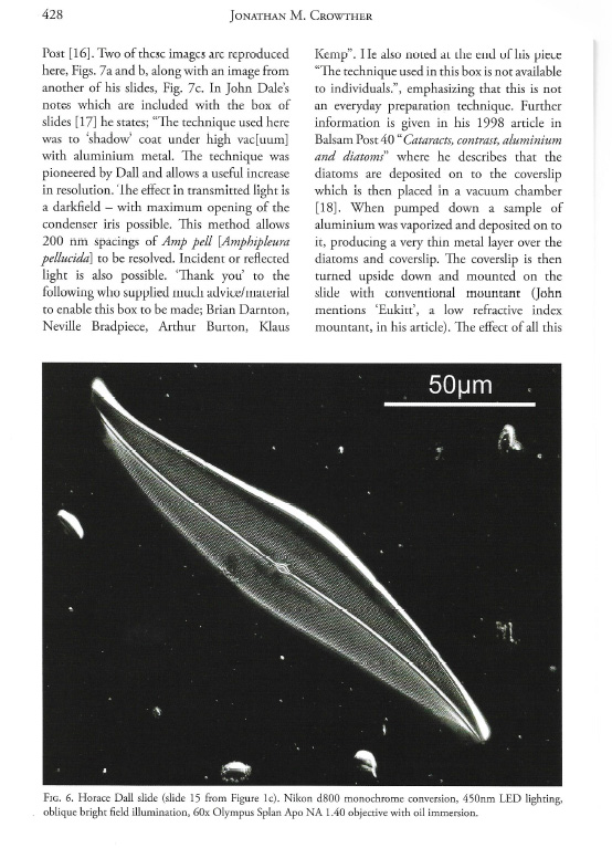
Latest publication – Microscopy of metal and metal oxide coated diatoms

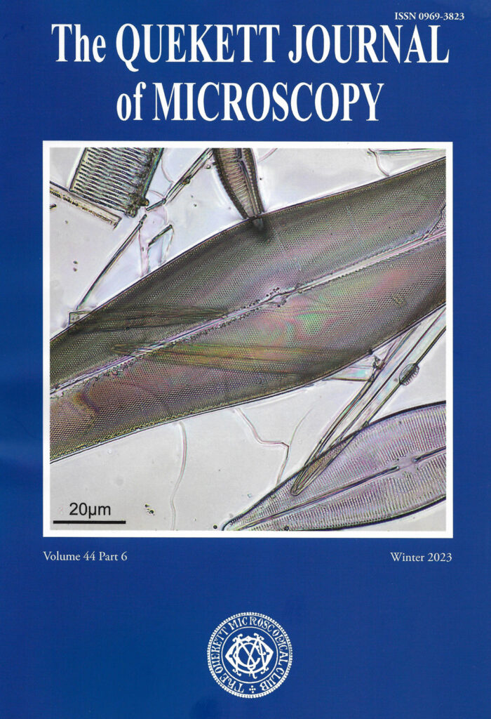
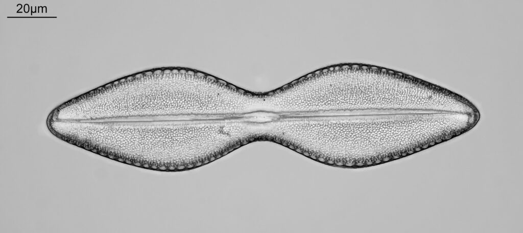
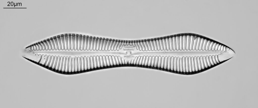
This is a nice way to end 2023 – a new publication of an article which was a fairly major piece of work this year. This one is the about the use of metal and metal oxide coating for diatom microscopy, and looks at the slides and techniques of making them from the very earliest examples to the modern day. It has been published in the Quekett Journal of Microscopy. I even managed to get one of my images as the cover art of this edition.

Shown below are some extracts from the article and the cover page.

Please excuse the relatively poor quality of the scans, my home scanner is not the most up to date. The hard copy in the Journal is much better.

It was as pleasure working on this one, as I find the approaches used to improve diatom visibility fascinating. Sourcing the slides was the biggest challenge as these are unusual items, but I wanted to write something to show the wider world some of what these amazing slide makers have produced.

More to come in 2024 as I have a few papers already in the pipeline. As always, thanks for reading, and if you’d like to know more about my work I can be reached here.
All the best for 2024 everyone!!!

Microscopy – Various diatoms, seasons greetings and 2024 plans

Firstly, seasons greetings to you all. I wish you the best for Christmas and the New Year. Today’s post is a chance for me to share some of my recent microscope images and also my thoughts on where my research will take me in 2024.

I am pretty certain that most of you come here for the pictures (rather than my deep and meaningful conversation), so lets jump straight into the images. All of these were done on my UV modified Olympus BHB microscope, but mainly with 450nm light rather than UV (easier to use with some of the slides). Camera for these was a monochrome converted Nikon d850 from MaxMax, and stacking was done using Zerene. Images have been reduced in resolution for sharing here. First up, a favourite of mine and a diatom that I’ve been on the look out for an intact copy of for a while – Hydrosilicon mitra, imaged using a 63x Leitz Pl Apo NA 1.40 objective and oil immersion, with oblique lighting.

Hydrosilicon mitra

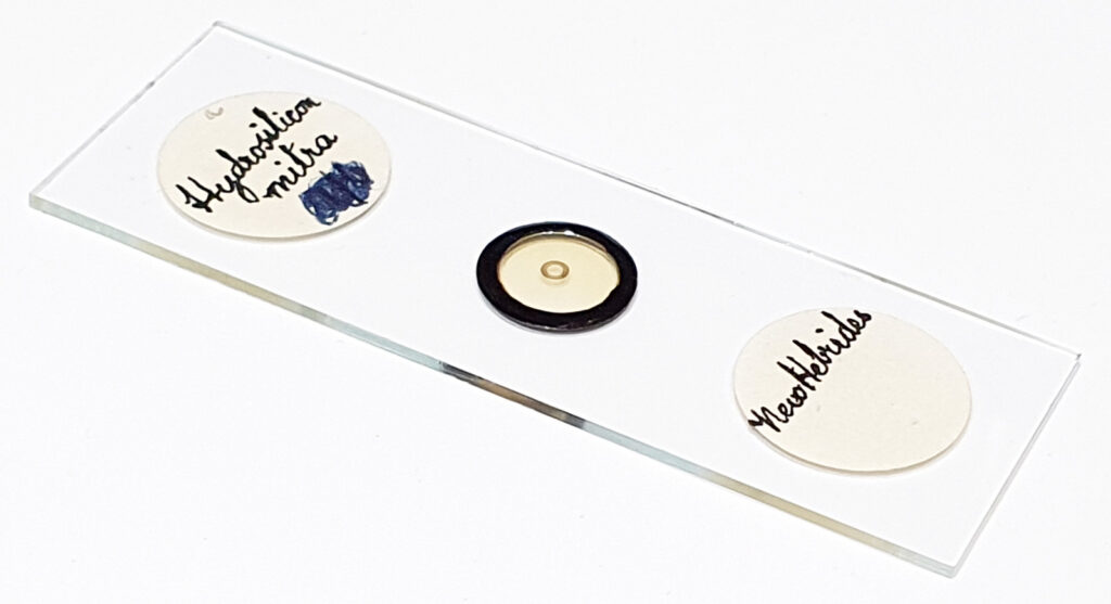
And the slide.

Hydrosilicon mitra slide

I believe the maker for this is JA Long, and there is a single example on the slide. Location for it – New Hebrides. The image was quite low contrast, as the mountant didn’t look like a high refractive index (RI) one. I have a couple of examples of this diatom but they have much more damage than this one does. I suspect this is a particularly delicate one to mount. A very unusual looking diatom.

Next up is Porodiscus hirsutus from an Oamaru strew slide by Mike Samworth. Again imaged using a 63x Leitz Pl Apo NA 1.40 objective with oil immersion, this time with bright field illumination.

Porodiscus hirsutus

And the slide.

Porodiscus hirsutus slide

A fabulous looking diatom with lots of interesting structures in it.

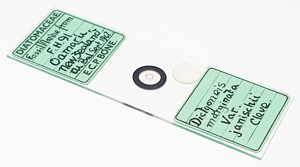
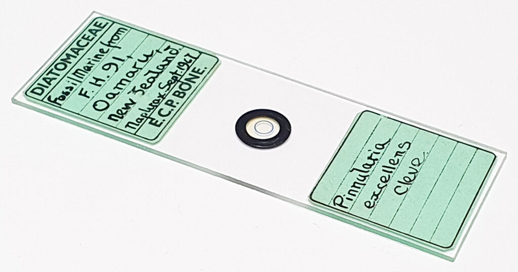
Next up two slides by ECP Bone. Both using the 63x Leitz Pl Apo NA objective with oblique lighting.

Dictyoneis marginata var. janischii
Pinnularia lobata

And the two slides.

Dictyoneis marginata var. janischii slide
Pinnularia lobata slide

Both of these are well made slides, each with a single example from Oamaru. Note that the Pinnularia lobata slide actually has the name Pinnularia excellens. This looks to be an error as the diatom looks more like the former than the latter (this happens more than you might expect due to the difficulties in identifying diatom species and the changes that have occurred with the names over the years).

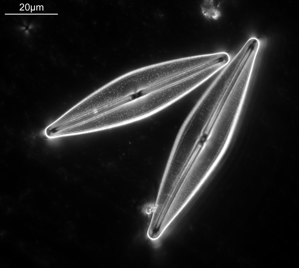
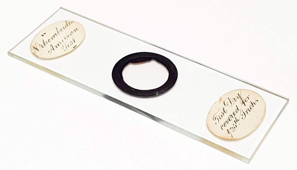
Finally for today an image from a Watson ‘Amichian test slide’ using darkground illumination (Reichert Neo 1.42/1.18 darkground condenser and 100x Leitz Pl Apo NA 1.32-0.60 condenser, both with oil immersion). For this one I went into the UV and used 365nm light.

Watson ‘Amician test slide’

And the slide.

Watson ‘Amician test slide’

The ‘Amician test’ is a test for resolution, and uses a Navicula diatom. However it is quite a mysterious test, and although this one says Navicula rhomboides, there is come controversy of which species was used for Amici’s original slide. Perhaps this will be something to write more about in the new year. Which leads me neatly on to the plans for 2024……

I like to have a few papers or articles which I am working on at any one time. A couple of papers are already planned for 2024. Firstly, some initial results on a new darkground condenser I’ve been working on. It is early days, but might be a neat idea for my UV darkground imaging where there are limited options available to buy (and by limited I mean as rare as hens teeth). Also building on an article I’ve recently written for the Quekett Journal of Microscopy on metal and metal oxide coated diatom slides, which looks at examples from their inception to now, I’d like to do a similar one from slides made using Realgar (a high RI material based on Arsenic Sulphide). This one needs a bit more thought, and ideally I need to get access to a 2d or 3d confocal Raman mapping device for a day to take a look at a slide in more detail, and try and understand more about what is going on chemically in the mountant. A work in progress.

Given I am now building up quite a catalogue of high resolution diatom images (the ones I have shared on this site are generally low resolution to I don’t exceed my storage capability) I’ve had a couple of people ask me if I am going to write a book or do something else with them. I’d like to do a book, but I know how much work they are. I think a mix of high res photos and some details on the slides, but combined with some essays on microscopy would be a good thing to do – a mix of art and science, and along the style of some of the early microscopists. Perhaps this can be my legacy to the microscopy world.

Anyway, as always, thanks for reading, and all the best for Christmas and 2024 whatever you are up to. If you’d like to know more about my work, I can be reached here.
Microscopy – Diatoms from Toome Bridge, Ireland

You can find find diatoms pretty much anywhere, but the range present at one location can be very different to other places. Toome Bridge (now called Toome or Toomebridge) in Ireland is a place which seems to have an amazing array of photogenic diatom species in the diatomite that is mined there. I have a couple of strew slides from that location and today’s post will show some images from one of those.

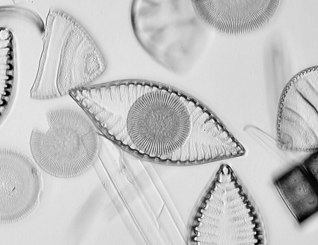
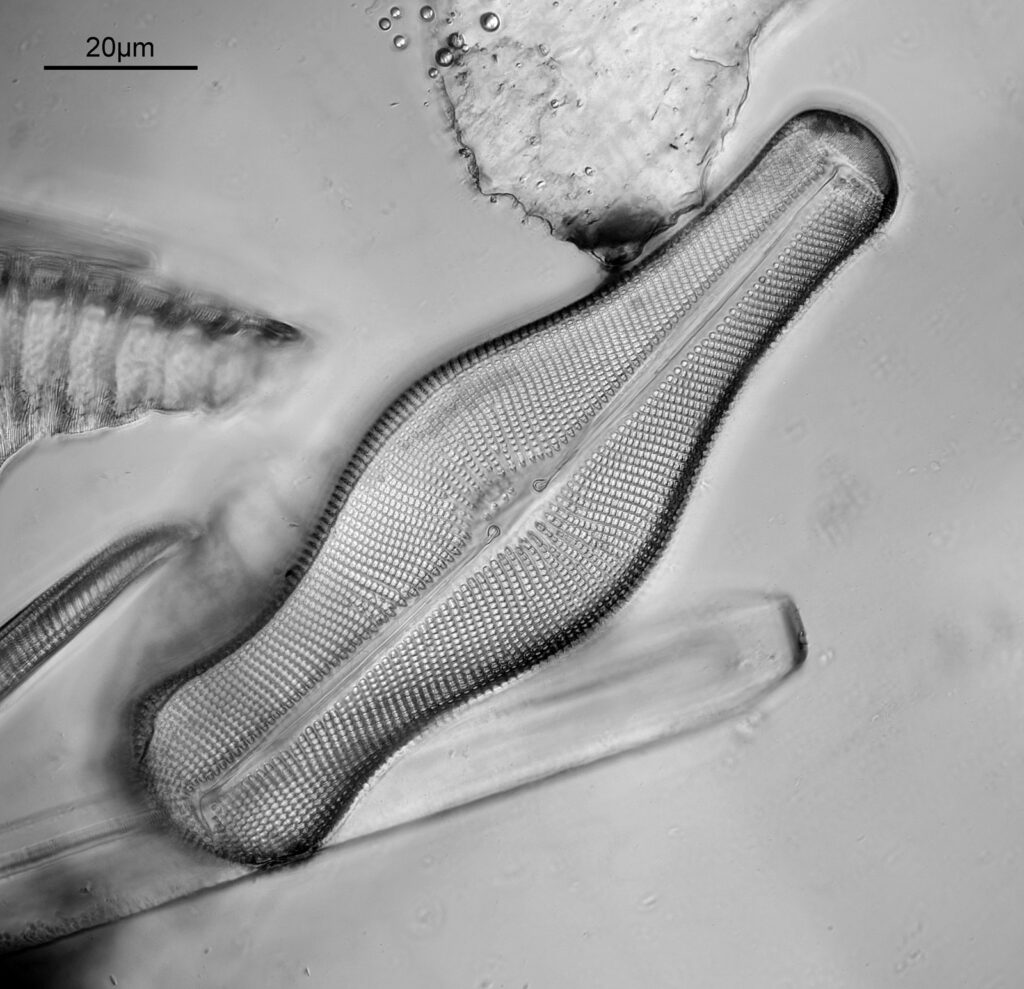
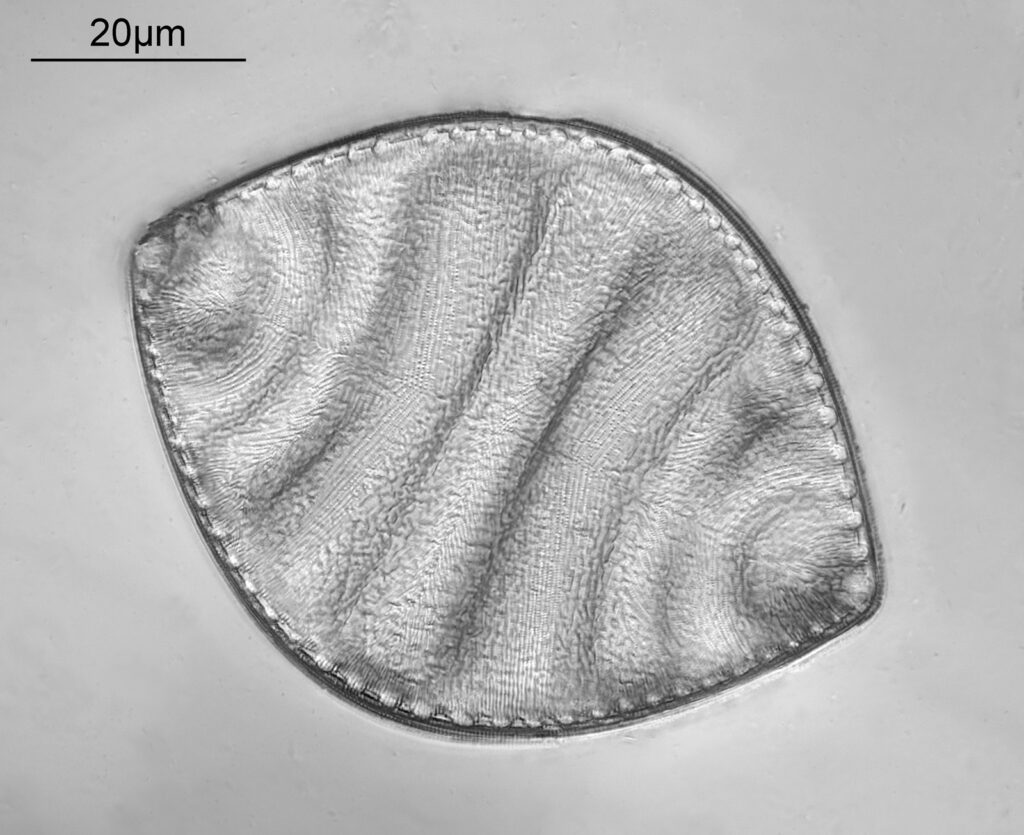
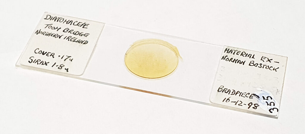
This slide was made by Neville Bradpiece, with diatoms mounted in Sirax. Apologies, but I don’t have names for all of these different species, but I have added names where I can. Some of the images have been reduced in resolution for sharing here. To start with, the eye…..

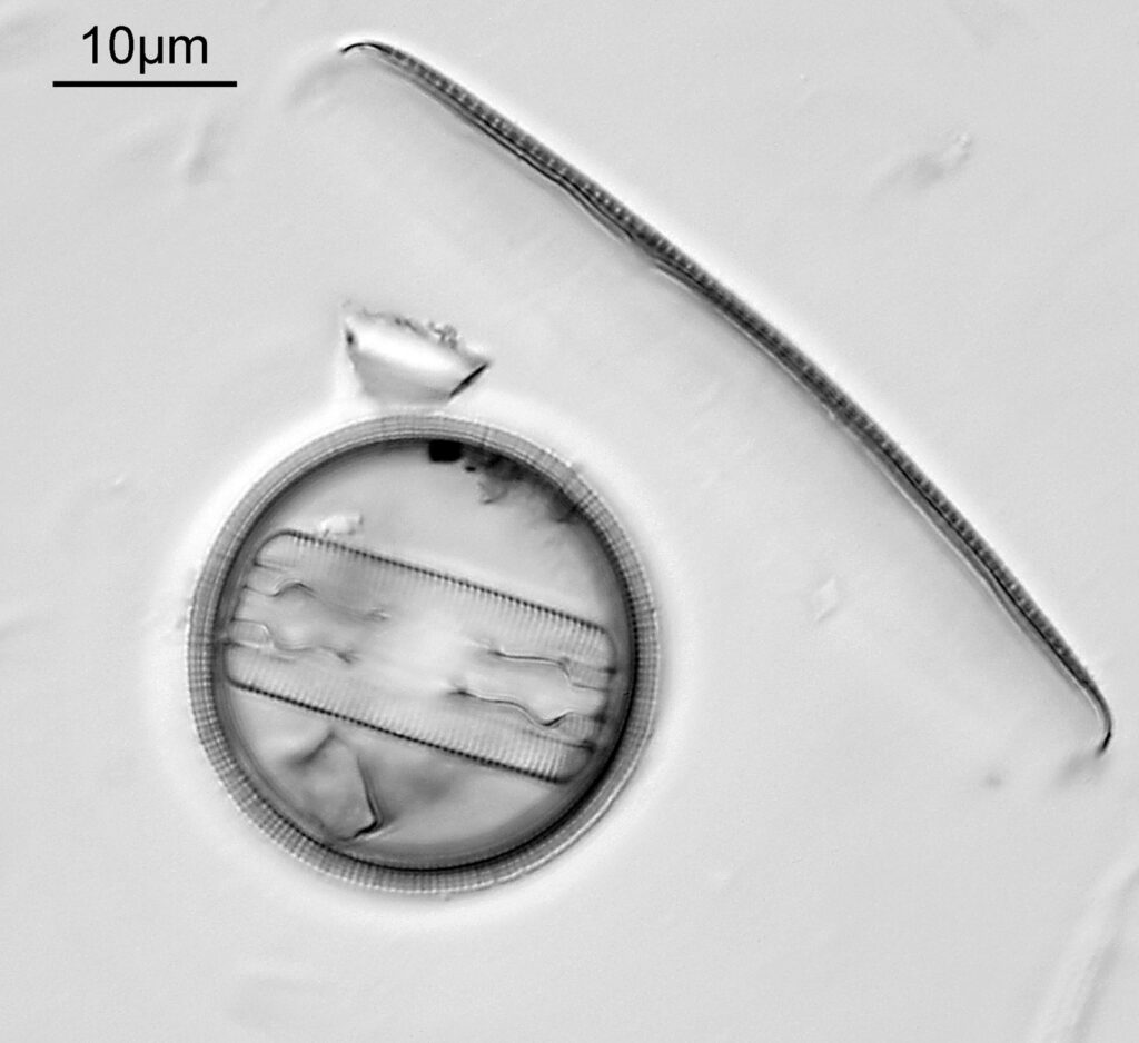
The image above has two diatoms in the centre, a circular one on top of a lens shaped one. This reminds me of an eye looking back at me through the microscope. Next is a tiny one, being only about 20 microns across.

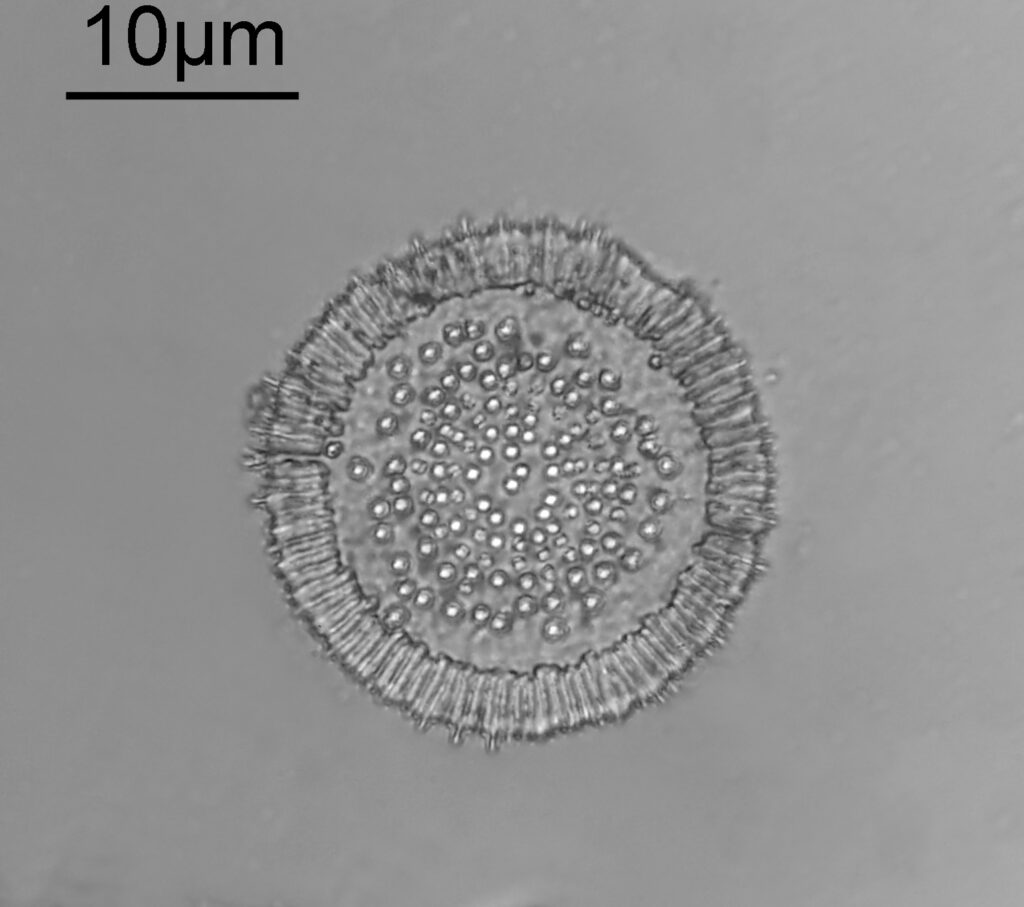
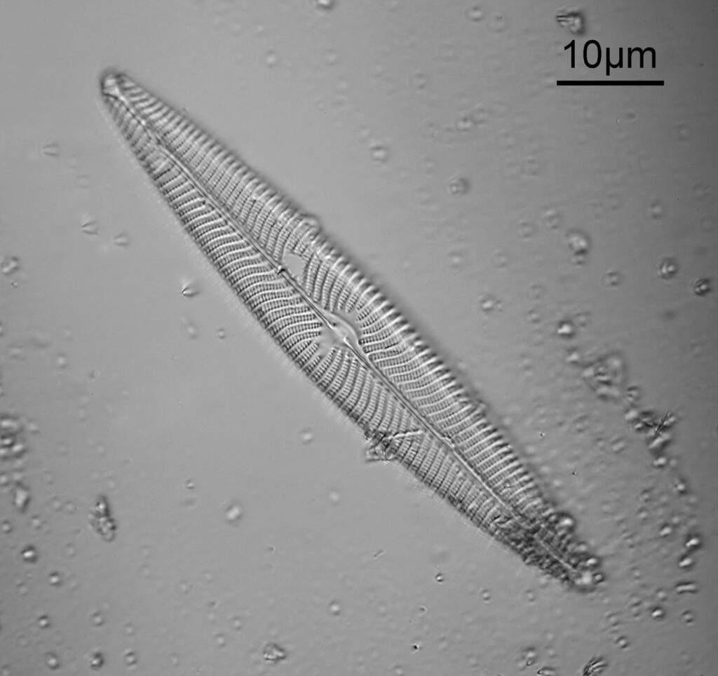
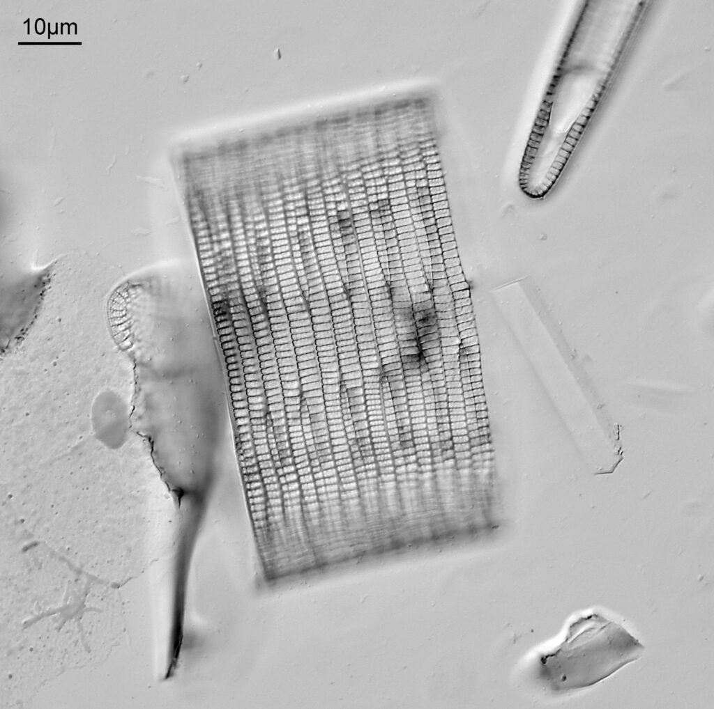
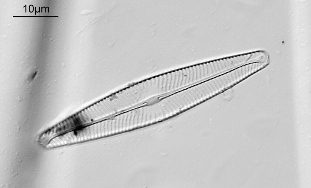
This one was tiny, being only about 20 microns across. Next is I think Navicula radiosa. I was using 365nm UV illumination for this slide, and a 63x Leitz Pl Apo objective with an NA of 1.40. Hence very high resolution.

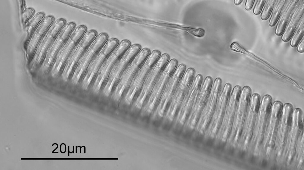
Below is part of a Pinnularia.

Going in close to this I got the following.

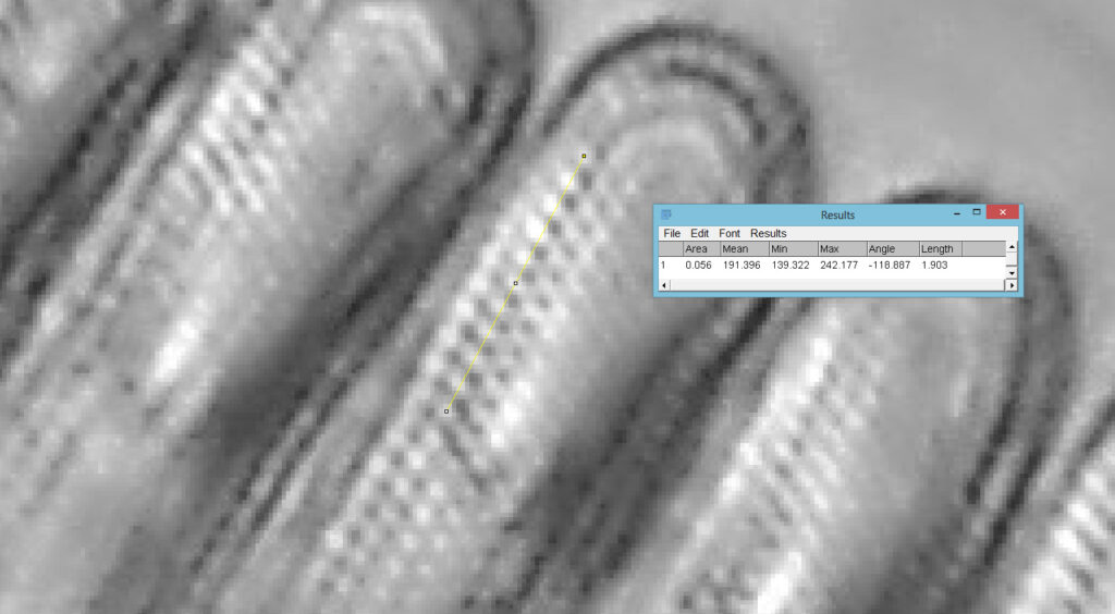
Based on the analysis in ImageJ, the spacing of the poroids here is about 190nm. Theoretical resolution with 365nm light and the NA 1.4 objective is about 130nm so this could well be correct, and it matches with SEM data I have seen for this species.

The one below is a Gomphomena.

The last one from this slide is a Cymatopleura elliptica.

Here’s the slide.

This slide caught my eye as it uses the mountant Sirax, and it mentions the very high refractive index it has (1.8). Sirax seems to be good for UV imaging as it is a high RI mountant which does not block the UV, hence I can get good contrast AND good resolution when using 365nm light. Note yet another spelling on the label – this time Toom Bridge. The images shared here scratched the surface of what was on the slide. I images upwards of a dozen different species from the slide, and that was not all of them. The more I looked the more I saw.

Strew slides are often overlooked in favour of slides with single species on them, but they can provide literally hundreds or thousands of diatoms to image, and are often cheaper than single species slides. This one cost me about £10, and I feel like I got an absolute bargain with it.

As always, thanks for reading and if you’d like to know more about any of my work, I can be reached here.

Microscopy – Northern Biological Supplies diatoms from Ramleh, Egypt

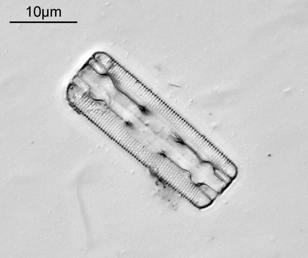
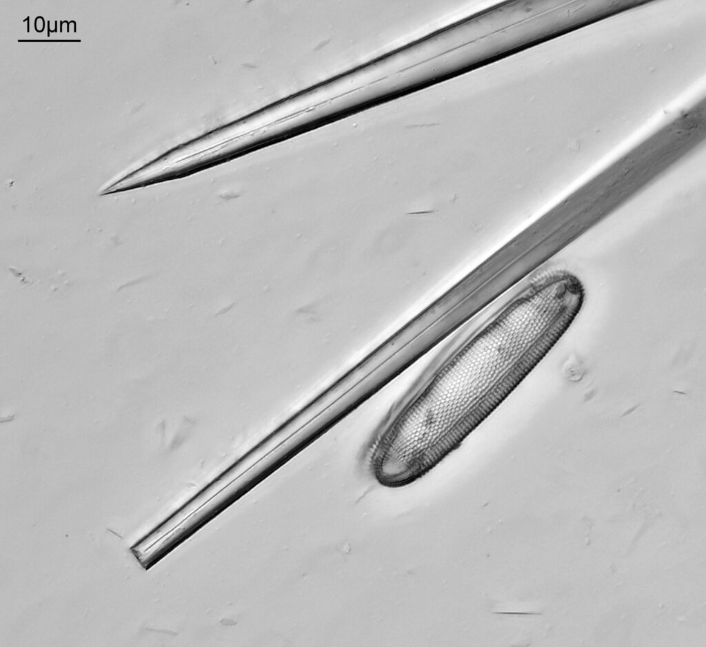
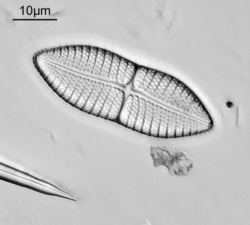
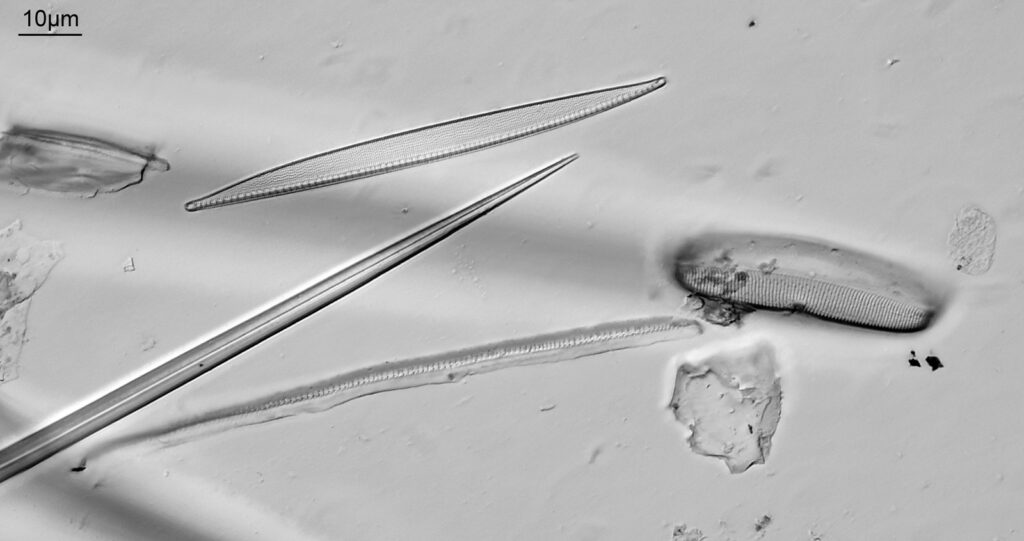
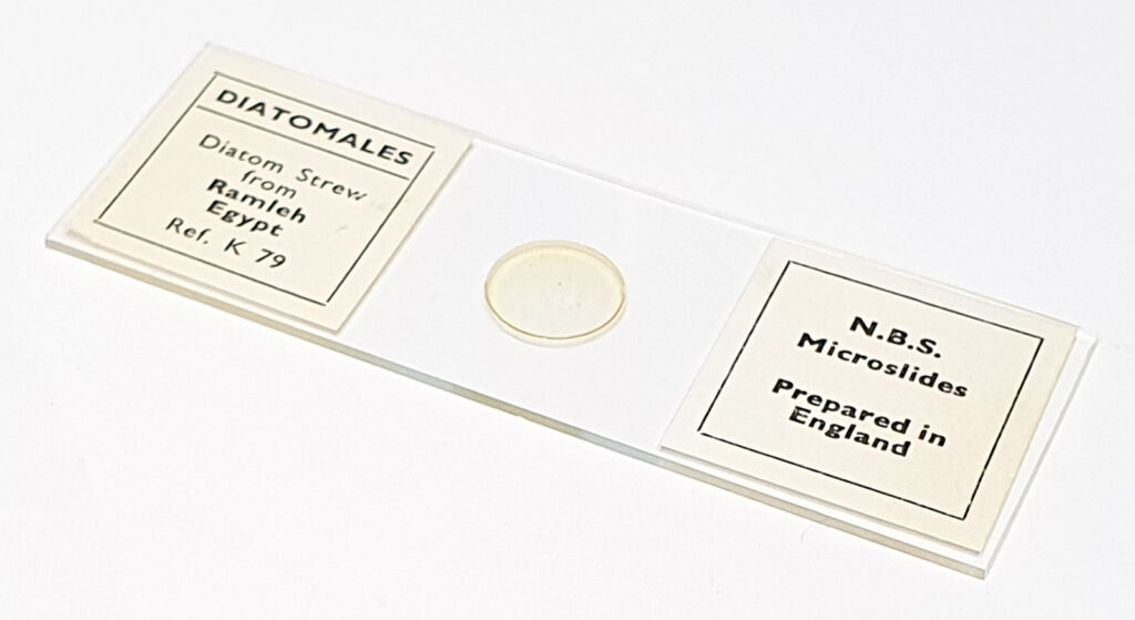
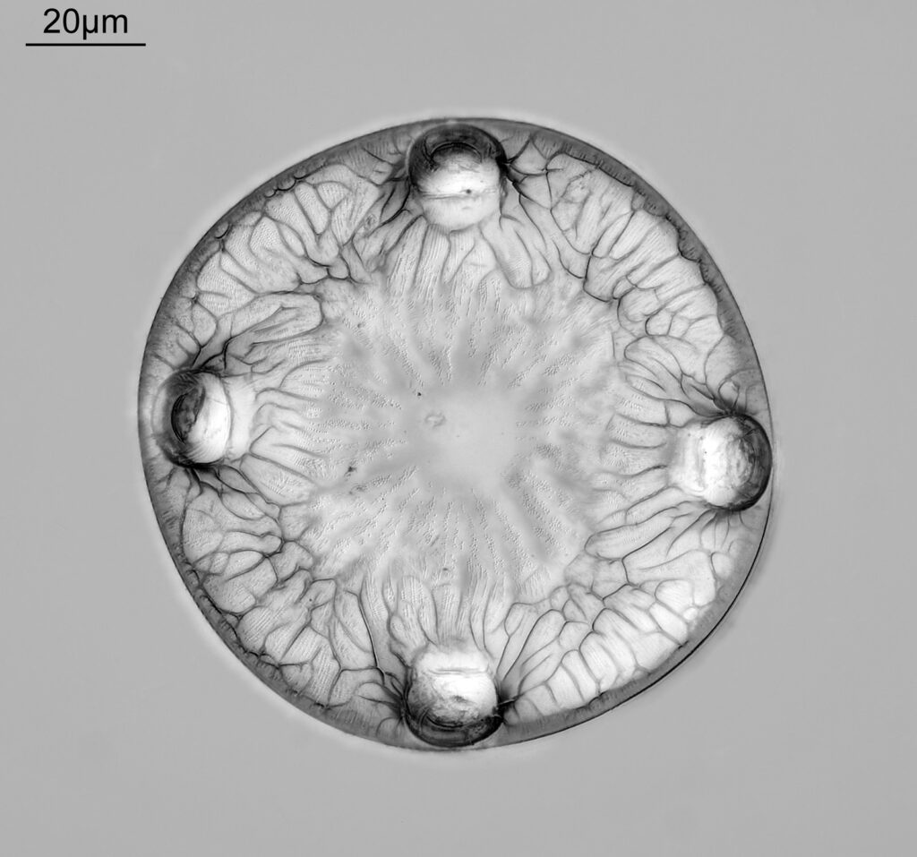
A few images to share today. These came from a slide made by Northern Biological Supplies (N.B.S.) and are from Ramleh, Egypt. N.B.S. slides tend to be well made and nice to image, and are worth looking for if you do not have some. These images were taken using my modified Olympus BHB microscope with 450nm LED light. A 63x Leitz Pl Apo NA 1.4 objective was used with oil immersion. Olympus Aplanat Achromat condenser, oil immersion, slightly oblique. 2.5x Nikon CF PL photoeyepiece. Monochrome converted Nikon d850 camera. Some images stacked using Zerene, others are just single photos.

There are a wide range of diatom species on the slide, and some spicules as well. Enough chat, here are the images.

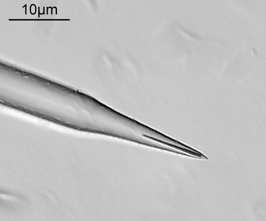
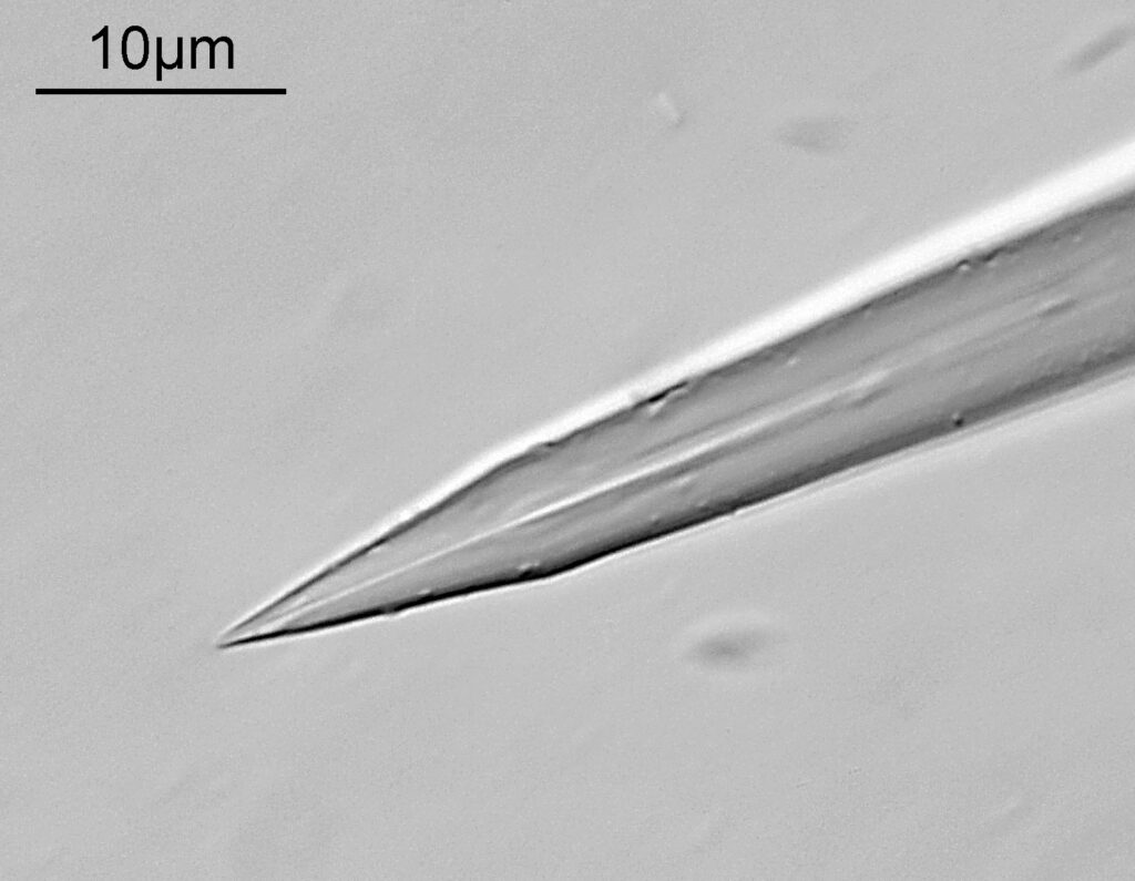
The last couple of images are closeups of the ends of a couple of spicules. Very sharp….. And the slide itself.

Strew slides are often overlooked by collectors and microscopists in favour of single species slides. However they can offer a wide range of subjects, and in some cases nice intact specimens (some strews are better than others for that) and generally cost less than the single species slides.

I hope you enjoy the images, and if you’d like to know more about my work, I can be reached here.
Imaging – the importance of clean camera sensors

Today I’m going to talk about dirt. Filth. Detritus. Contamination. Call it what you want, but a dirty camera sensor can have a big impact on the imaging process, requiring a lot of time to deal with in the processing stage. With microscopy this is potentially even worse than with normal photography as the objectives mean that the effective aperture of the lens is tiny, making dirt on the sensor even more obvious. Also as the wavelength of light goes down, the resolution goes up, so shorter wavelength light further compounds the problem. This impacts my UV work. I normally avoid cleaning camera filters, as I’ve never had much luck with it, and there is the chance that you can make it much worse , however recently I’ve found myself having to spend more and more time on removing the dirt from my microscope images in Photoshop. So I took the plunge and set about getting the right kit to clean my monochrome converted Nikon d850.

While perhaps not as interesting to the wider audience as my normal microscopy posts, this is an important topic. In ‘manager speak’ time is money, and if I spend an extra 20-30mins processing every diatom image to remove the dirt in Photoshop, that time all adds up. As my diatom imaging is not funded, this is not directly costing me money, but the time lost is something I could be doing something else with. Before we start, a nice diatom image from a slide by SH Meakin (an Auliscus speciosus). This was done after cleaning the sensor, and was so much easier to work on.

Auliscus speciosus diatom by SH Meakin.

To do my cleaning I needed some kit. And yes, I have cleaned camera sensors before with a cotton bud and breathing on the sensor, but the right kit makes things a lot easier. I contacted Just Clean here in the UK, and after watching their videos on how to clean a sensor, I realised there were three main steps to consider – removal of the loose dirt with a blower, using a Dustaid to mechanically remove dirt on the surface of the sensor coverglass and then a swab with a cleaning solution as the final step. I called them up and they were very helpful, checking what cameras I had (and what sized sensors) and recommending the right cleaning products.

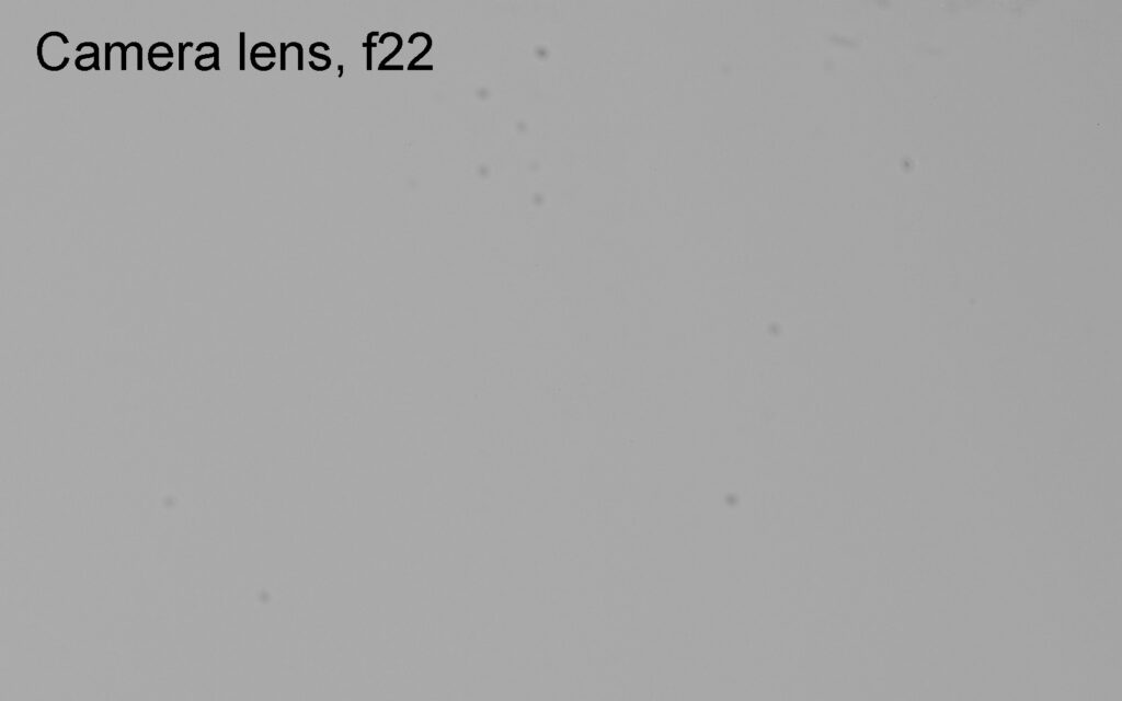
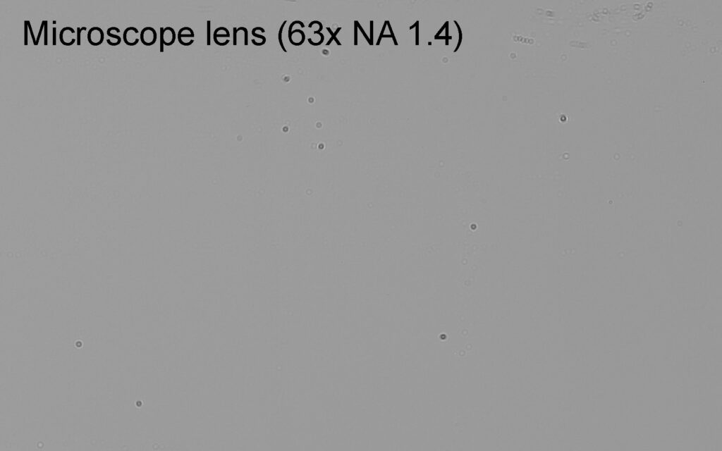
Before cleaning I did an image of a white screen with a normal camera lens, at f22 (75mm on a zoom), and this a crop of the top right off the image at original pixel resolution.

The fuzzy blobs are dirt on the sensor, and while they are visible they are not hugely distracting with this lens and f stop. Next was the same camera, still before cleaning, but on the microscope using 450nm light, and a 63x Leitz Pl Apo NA 1.4 objective, which is one I often use for my diatom imaging work.

With the camera on the microscope, the blobs become much more well defined, and there are a lot more of them which are obvious.

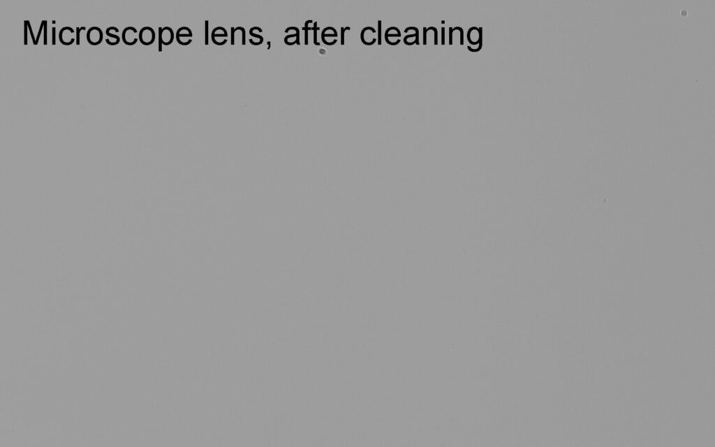
Then the sensor was cleaned (blower, Dustaid and swab), and the camera put back on the microscope and another image was taken with the same setup.

As can be seen, cleaning has removed the vast majority of the dirt as there are now far fewer blobs on the image.

I think where some people can struggle with cleaning is ‘how clean is clean?’. While it may be nice to think that cleaning removes all the dirt that just isn’t realistic. It’ll always be a case of managing expectations – what is clean enough? For me if I can remove the vast majority of the dirt from the sensor I am not having to spend time on Photoshop touching up every image, so for me this was now clean enough. Will I clean the sensor every week? No. Will I keep an eye on it and clean it again when it gets annoying enough to worry about? Yes.

Unfortunately, despite our best efforts camera sensors will get dirty. Cleaning them can be done at home if you feel confident enough to take it on. If not then there are places which offer it as a service, but keep in mind, that after a clean if a camera then gets knocked around in the shipping process, that can liberate more dust from the inside which can end up back on the sensor. So you have to be realistic about how clean it would be when you get it back.

As always, thanks for reading, and if you’d like to know more about my work, I can be reached here.
Microscopy – Bausch and Lomb 53x UV reflecting objective imaging of diatoms

Occasionally I like to have a play with some of the unusual historical UV microscope equipment I’ve been fortunate to be able to find, and see whether it can still be used to create images on my Olympus BHB. Last night I got a Bausch and Lomb 53x NA 0.72 reflecting objective out of its box and put it on the microscope. Today’s post shares some images from it and a bit about the objective itself.

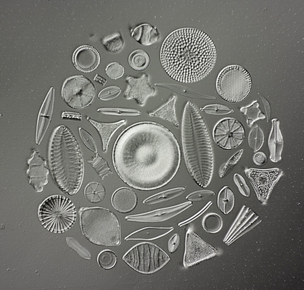
To start with here are a couple of images using the objective (resolution reduced for sharing here). Both images were done using 450nm light.

Diatom using the Bausch and Lomb 53x UV reflecting objective.

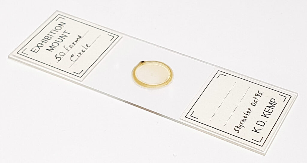
The images were relatively low contrast compared with the objectives I normally use, but this is not unexpected for reflecting objectives. The slide was a 50 form diatom one by Klaus Kemp, shown below.

Klaus Kemp 50 form slide (using a 10x objective).
Klaus Kemp 50 form slide.

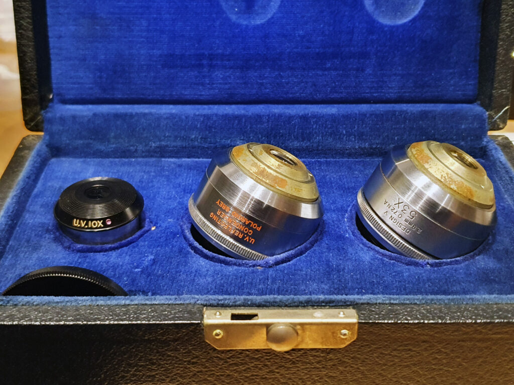
The Bausch and Lomb (B&L) UV reflecting objectives are unusual and rare. I’ve seen them discussed in a 1958 B&L catalogue, and there were two types – a 53x NA 0.72 and a 94x NA 1.00 – each along with matching reflecting condensers, and 3.5x and 10x UV eyepieces. The reflecting objectives and condensers combined quartz and calcium fluoride refractive elements along with mirrors. The 10x eyepiece was quartz, and the 3.5x quartz and calcium fluoride. I’d assumed these were unicorns and would never see them in the flesh, and then I managed to track down a boxed set of the 53x objective and condenser and 10x eyepiece and jumped on it. Here it is.

Bausch and Lomb 53x UV reflecting objective and condenser and 10x UV eyepiece.

Cosmetically they have a little corrosion on the outside of their housings but optically they looked pretty good. Measurement of the UV transmission through them did indeed show there was no normal glass in them, and they transmitted light down to less than 280nm. Interestingly despite the box being B&L, on the back of the objectives it says ‘Polaroid Grey’. My assumption is that they have been made for B&L by Polaroid Grey, but that is an assumption.

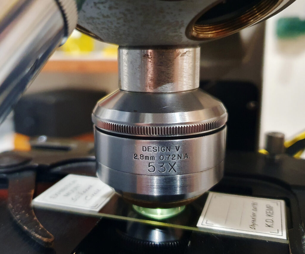
I mounted the objective on my microscope using a small RMS extension tube (the working distance is less than a 1mm with this objective, and the extension tube mean that I didn’t need to raise the stage too far). The eyepiece was placed where photoeyepiece normally goes. This is shown below.

53x B&L UV reflecting objective in place.
10x B&L UV eyepiece in place.

This was very much ‘suck it and see’ experiment. I just set it up and tried it out, and was happy to get some images (image stacking was done using Zerene). Depth of field was like razor blade, and initial focusing was fun, especially as working distance was about 0.7mm. There is some dirt in the eyepiece, so I need to take that apart and clean it. The objective can probably have the alignment optimized, but not by me. It was probably either not meant to be done after being set in the factory, or to be sent back to B&L for it to be done. I doubt if there is still anyone around would know how to do it, so that will be left alone. I did not use the matching reflecting condenser, as there is no way to mount that on my Olympus BHB microscope. 450nm light was used as this was more simple than setting up for UV source for the test, but this would be usable down to and below 300nm as it is UV transparent.

There are some fascinating historical optical items out there which would have been horrendously expensive to buy when they were originally made. While rare they do crop up occasionally for sale, and that allows people like me to try out a slice of history for my research. As always, thanks for reading, and if you’d like to know more about my work, I can be reached here.
Microscopy – The beauty of arranged diatom slides

The Victorians were masters of making arranged diatom slides, and a range of makes produced these from the late 1800s into the 1900s. While there are still some that have been making them recently – Klaus Kemp (now deceased) and Steve Beats, for example – these early examples of arranged slides remain as amazing works of art, and demonstrations of technical expertise. I’ve recently been fortunate enough to acquire a number of these slides and want to share some initial images from them.

The first is one by Watson and Sons Ltd, and is a circular group from St Peters, Hungary (resolution has been reduced for sharing here). I think this one dates to some time around the 1930s.

Watson and Sons circular diatom arrangement

And the slide.

Watson and Sons circular diatom arrangement slide

Imaging the whole circle was a bit of a challenge as it is about 3.5mm across. I ended up using a 3.2x Zeiss Semiplan NA 0.10 objective and removing the condenser from the microscope.

The next is an 80 form type slide by Möller.

80 form type slide by Möller.

This has examples of 80 different diatom species along with a tiny printed grid with the diatoms names. This is about 3mm across. The 2nd diatom from the left of the top row has moved and is now out of focus. This is not hugely uncommon in these old arrangements, but I still find it amazing how one of the diatoms can move through the mountant while all the others remain where they are. One of life’s mysteries…..
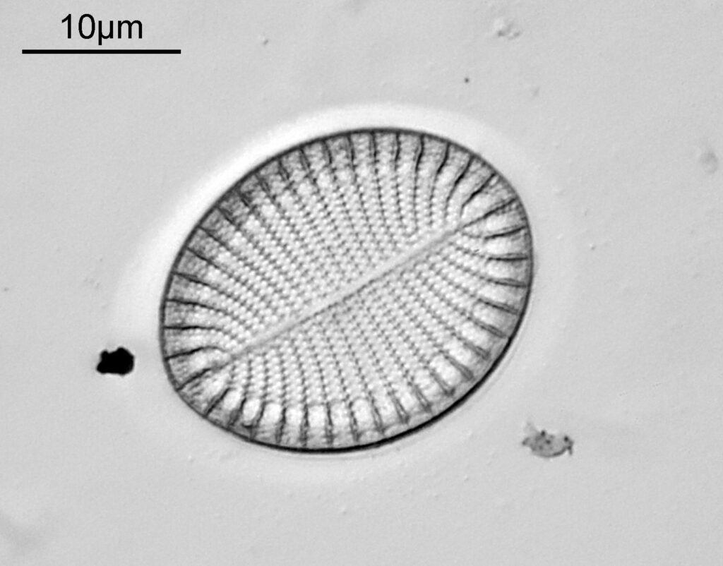
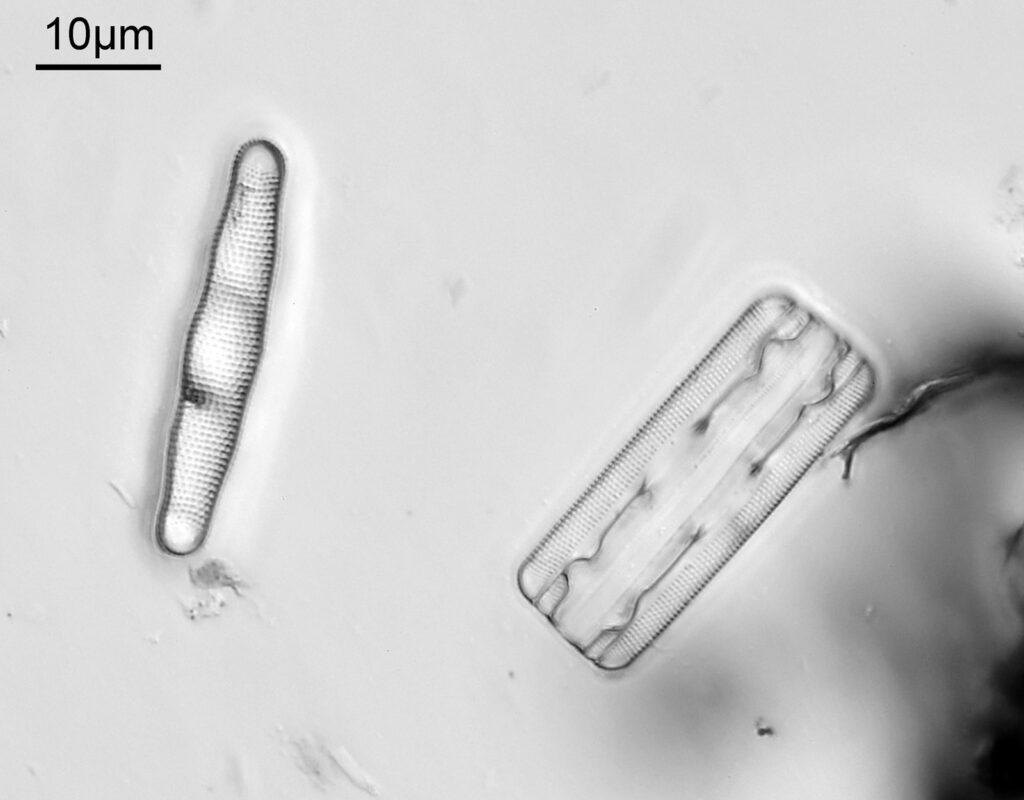
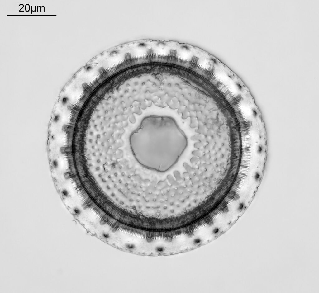
Below is an image of one the bottom left diatom – Stephanodiscus nigarae – and a close up on the writing.

The scale bar gives an example of just how small this writing is. From what I can tell the writing is on the underside of the coverslip, and then the diatoms are mounted below that (based on how the image changed as I altered the position of the stage).

Now, how to best share images of these slides in the future? Ideally I’d like to image and share them at high resolution, but my current website isn’t built for sharing many large images. I shall need to have a think about how best to deal with these slides.

As always. thanks for reading, and if you’d like to know more about my work I can be reached here.