
Firstly, seasons greetings to you all. I wish you the best for Christmas and the New Year. Today’s post is a chance for me to share some of my recent microscope images and also my thoughts on where my research will take me in 2024.
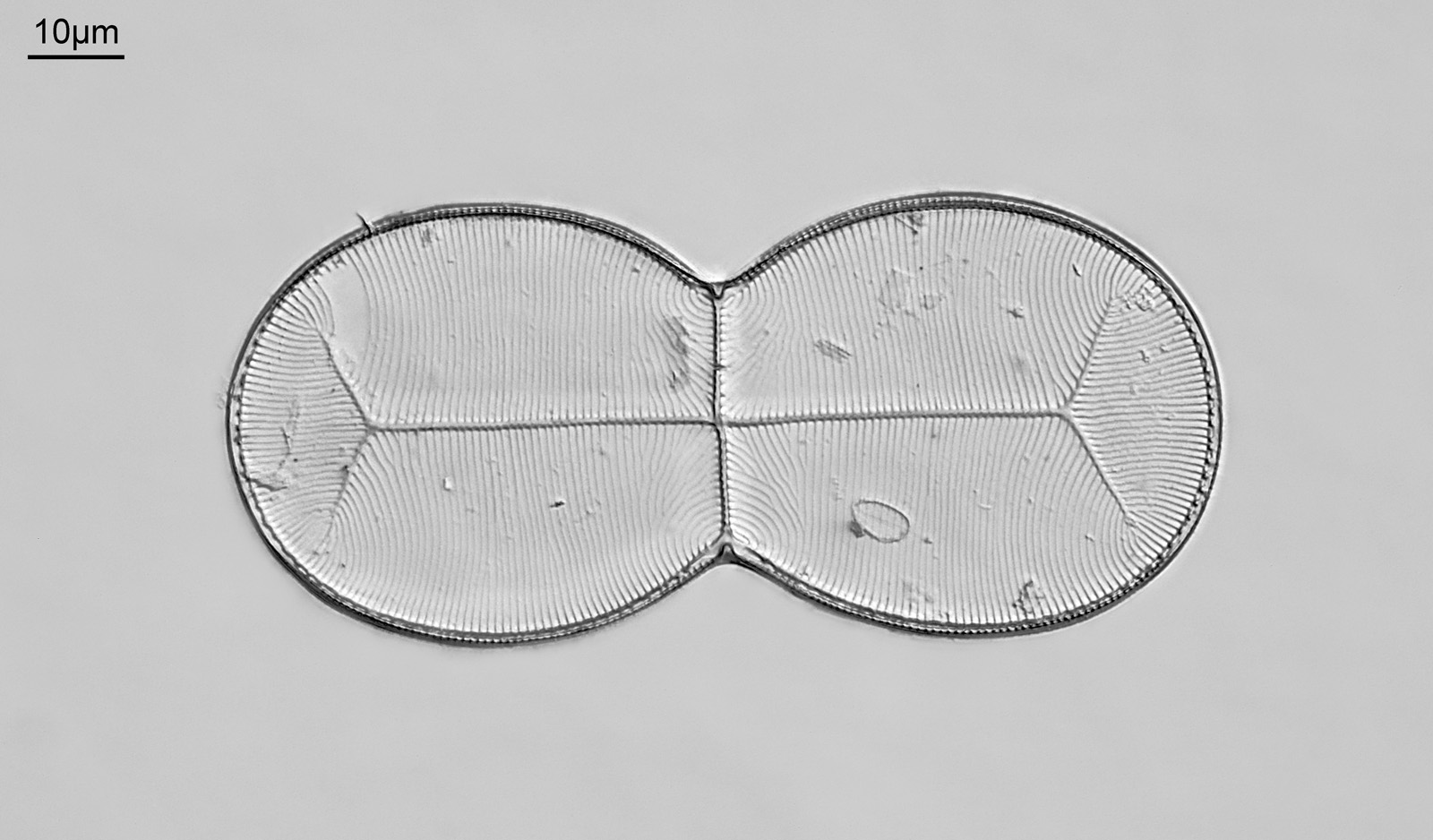
I am pretty certain that most of you come here for the pictures (rather than my deep and meaningful conversation), so lets jump straight into the images. All of these were done on my UV modified Olympus BHB microscope, but mainly with 450nm light rather than UV (easier to use with some of the slides). Camera for these was a monochrome converted Nikon d850 from MaxMax, and stacking was done using Zerene. Images have been reduced in resolution for sharing here. First up, a favourite of mine and a diatom that I’ve been on the look out for an intact copy of for a while – Hydrosilicon mitra, imaged using a 63x Leitz Pl Apo NA 1.40 objective and oil immersion, with oblique lighting.
And the slide.
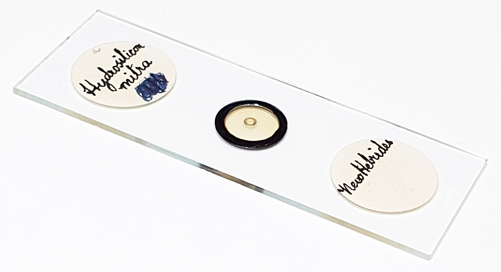
I believe the maker for this is JA Long, and there is a single example on the slide. Location for it – New Hebrides. The image was quite low contrast, as the mountant didn’t look like a high refractive index (RI) one. I have a couple of examples of this diatom but they have much more damage than this one does. I suspect this is a particularly delicate one to mount. A very unusual looking diatom.
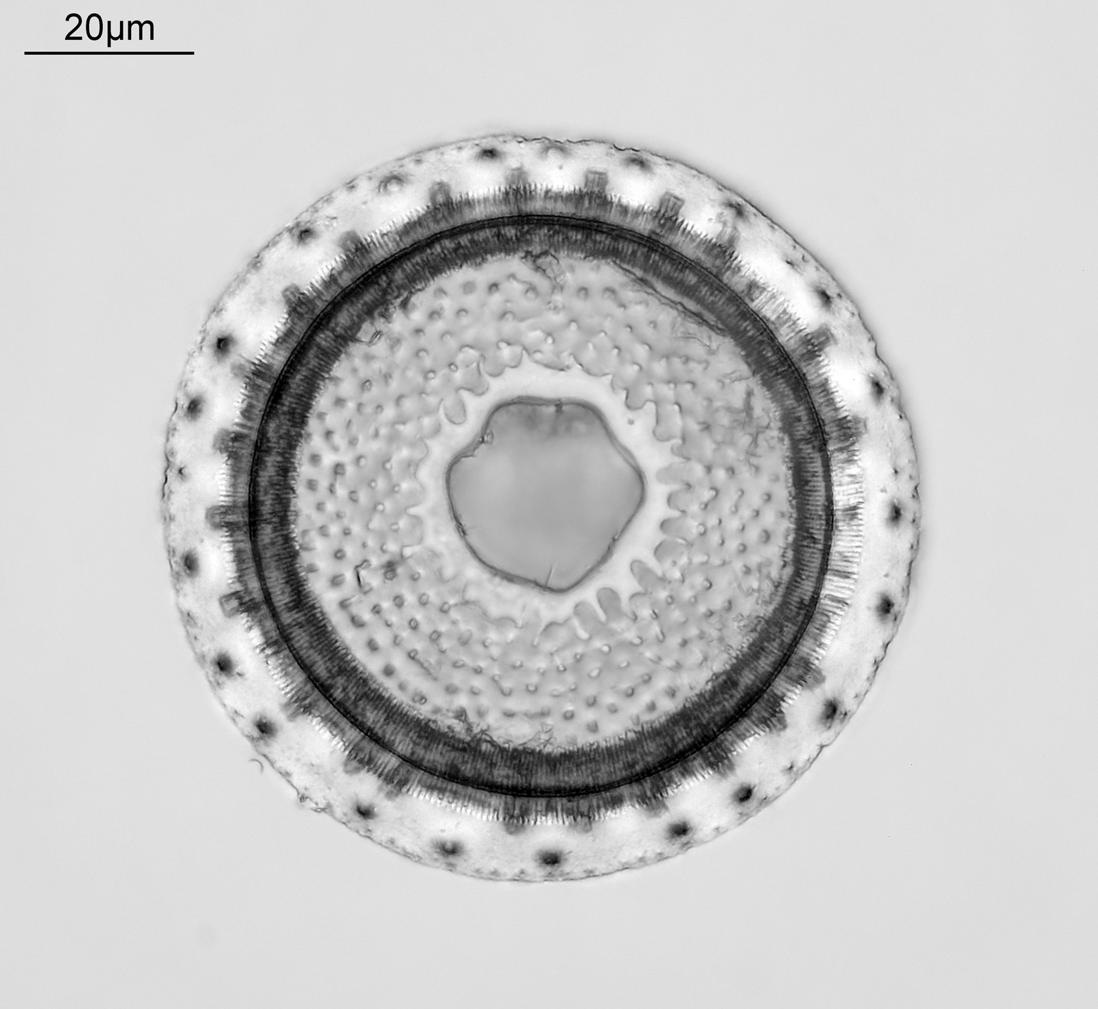
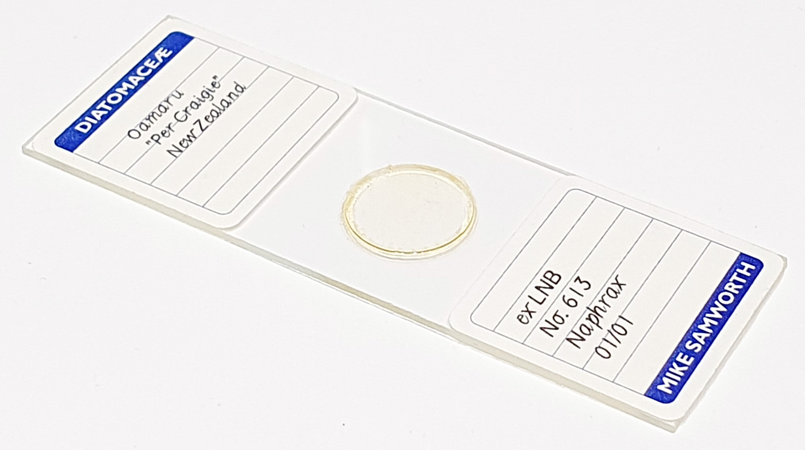
Next up is Porodiscus hirsutus from an Oamaru strew slide by Mike Samworth. Again imaged using a 63x Leitz Pl Apo NA 1.40 objective with oil immersion, this time with bright field illumination.
And the slide.
A fabulous looking diatom with lots of interesting structures in it.
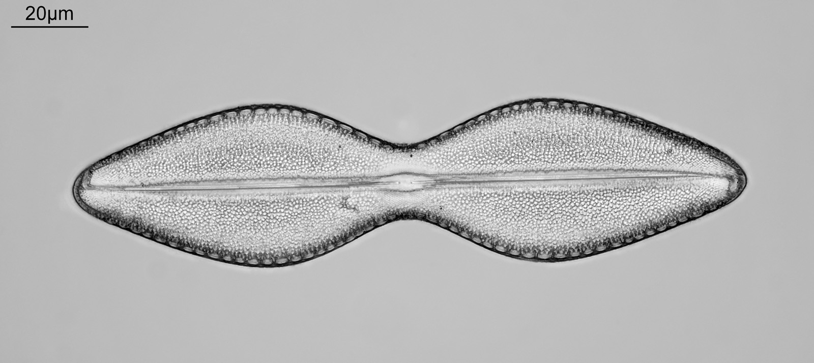
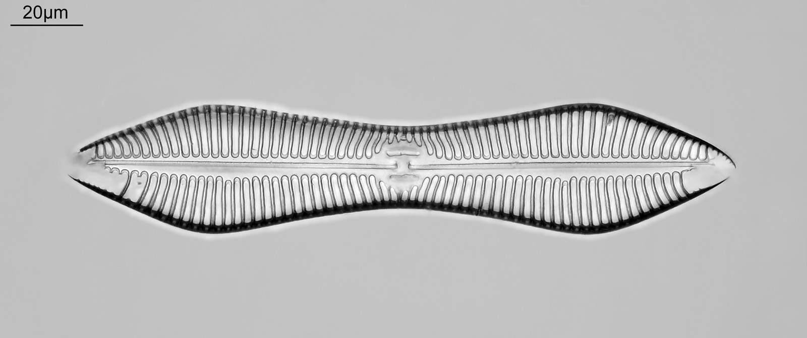
Next up two slides by ECP Bone. Both using the 63x Leitz Pl Apo NA objective with oblique lighting.
And the two slides.
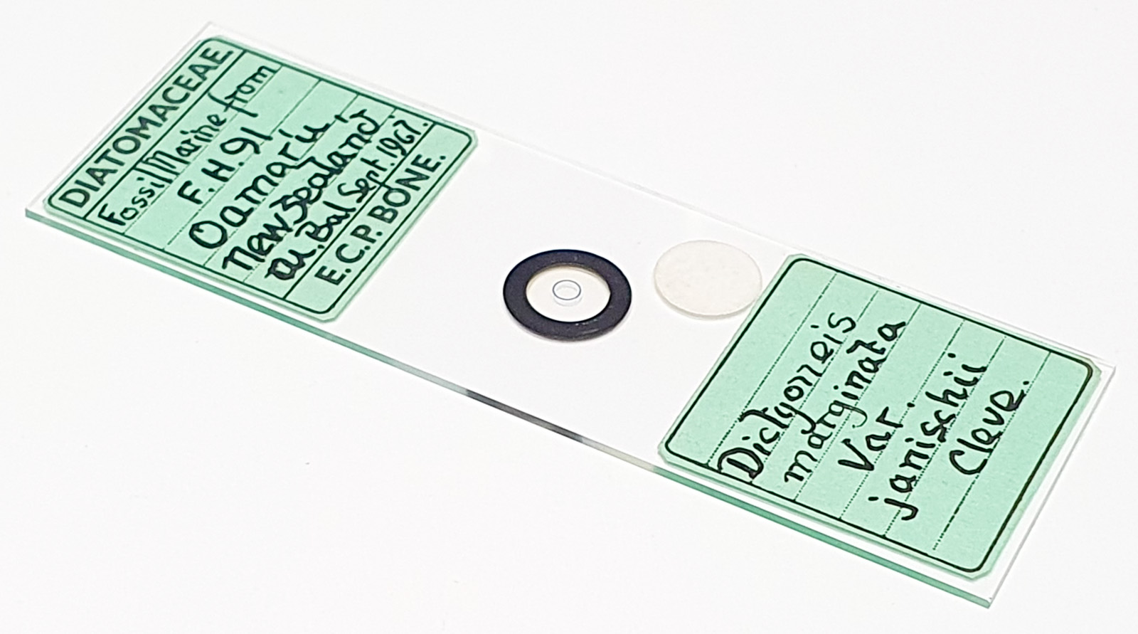
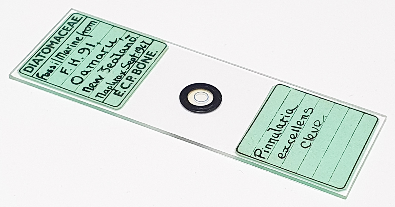
Both of these are well made slides, each with a single example from Oamaru. Note that the Pinnularia lobata slide actually has the name Pinnularia excellens. This looks to be an error as the diatom looks more like the former than the latter (this happens more than you might expect due to the difficulties in identifying diatom species and the changes that have occurred with the names over the years).
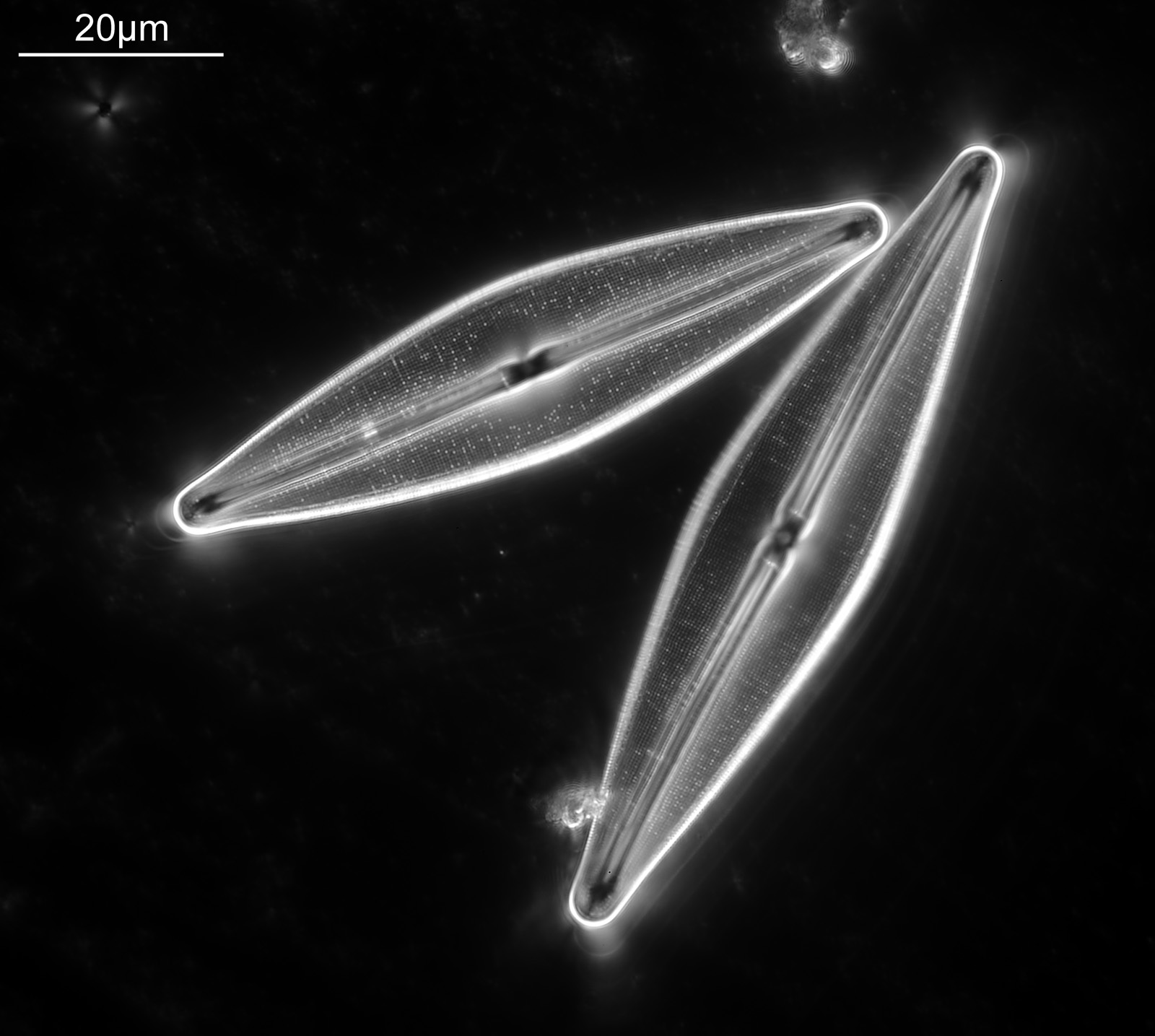
Finally for today an image from a Watson ‘Amichian test slide’ using darkground illumination (Reichert Neo 1.42/1.18 darkground condenser and 100x Leitz Pl Apo NA 1.32-0.60 condenser, both with oil immersion). For this one I went into the UV and used 365nm light.
And the slide.
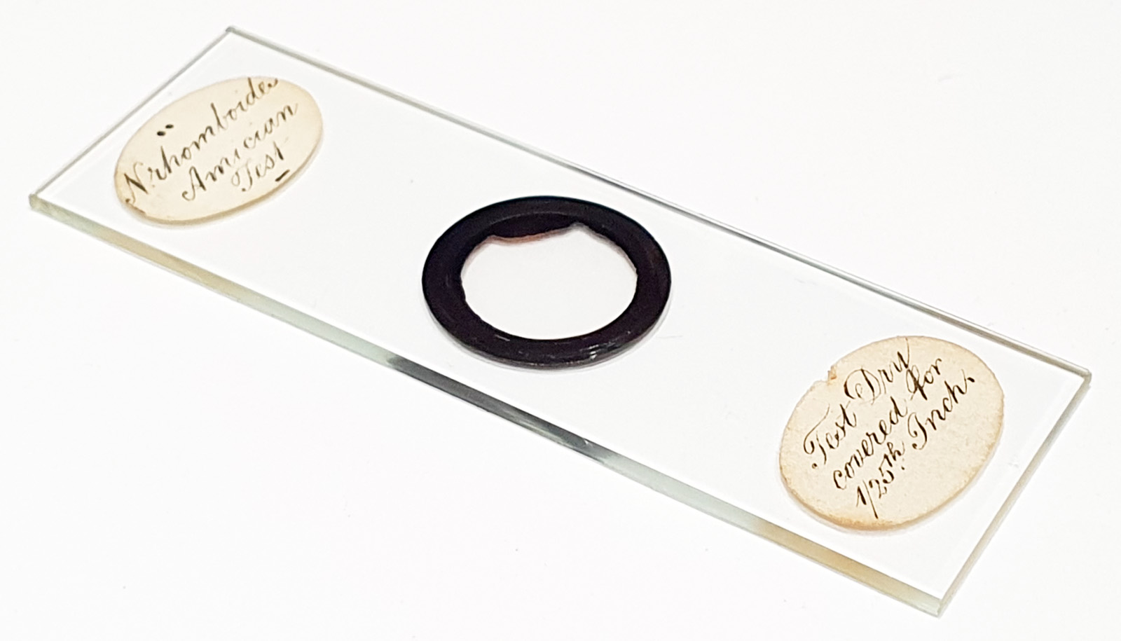
The ‘Amician test’ is a test for resolution, and uses a Navicula diatom. However it is quite a mysterious test, and although this one says Navicula rhomboides, there is come controversy of which species was used for Amici’s original slide. Perhaps this will be something to write more about in the new year. Which leads me neatly on to the plans for 2024……
I like to have a few papers or articles which I am working on at any one time. A couple of papers are already planned for 2024. Firstly, some initial results on a new darkground condenser I’ve been working on. It is early days, but might be a neat idea for my UV darkground imaging where there are limited options available to buy (and by limited I mean as rare as hens teeth). Also building on an article I’ve recently written for the Quekett Journal of Microscopy on metal and metal oxide coated diatom slides, which looks at examples from their inception to now, I’d like to do a similar one from slides made using Realgar (a high RI material based on Arsenic Sulphide). This one needs a bit more thought, and ideally I need to get access to a 2d or 3d confocal Raman mapping device for a day to take a look at a slide in more detail, and try and understand more about what is going on chemically in the mountant. A work in progress.
Given I am now building up quite a catalogue of high resolution diatom images (the ones I have shared on this site are generally low resolution to I don’t exceed my storage capability) I’ve had a couple of people ask me if I am going to write a book or do something else with them. I’d like to do a book, but I know how much work they are. I think a mix of high res photos and some details on the slides, but combined with some essays on microscopy would be a good thing to do – a mix of art and science, and along the style of some of the early microscopists. Perhaps this can be my legacy to the microscopy world.
Anyway, as always, thanks for reading, and all the best for Christmas and 2024 whatever you are up to. If you’d like to know more about my work, I can be reached here.