
This is a bit of technical update post, as it covers something I wasn’t aware of before, which may be of interest to other microscopists. A few days ago, I was doing some work with an old 170x Leitz Q NA 0.5 reflecting objective (see here) and noticed that when the aperture of the condenser was closed right down I got a darkfield image, with a black background. This got me wondering whether it would work with other reflecting objectives, and whether it was possible to use this technique to get darkfield images in the UVB region at 313nm (something I previously couldn’t attempt as I don’t have a quartz darkfield condenser). This post gives and update to that work.
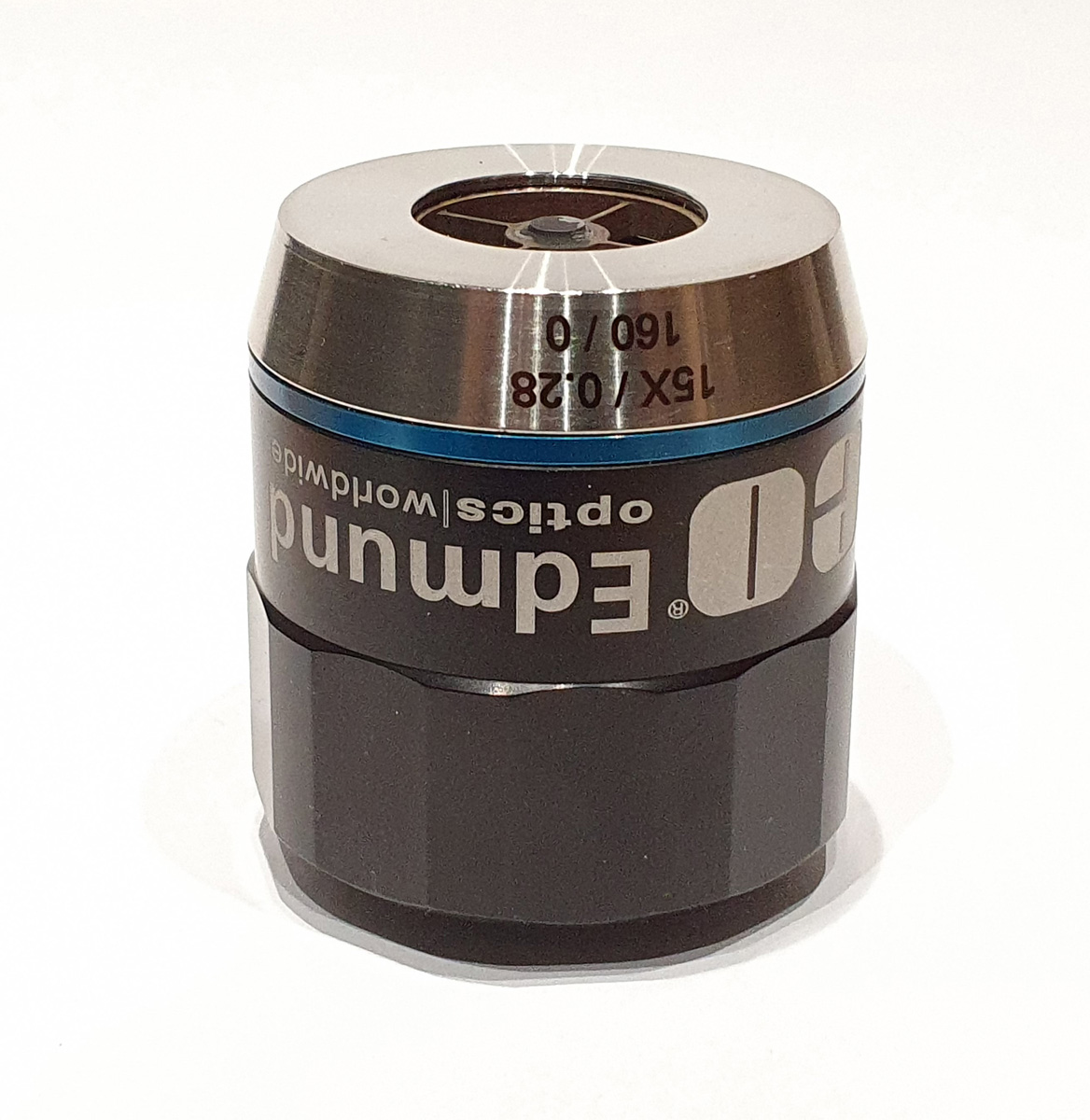
As a subject for imaging I chose the diatom slide which I’d had prepared using quartz slide and coverslip which I have been using for imaging at 313nm (examples here). Rather than use the 170x Leitz reflecting objective, which wasn’t designed for a microscope with a tube length like mine, I used a 15x Edmund Optics NA 0.28 reflecting objective. The condenser was my vintage Zeiss quartz one. With the condenser iris closed right down, imaging at 313nm, I got the following image of one of the diatoms on the slide.
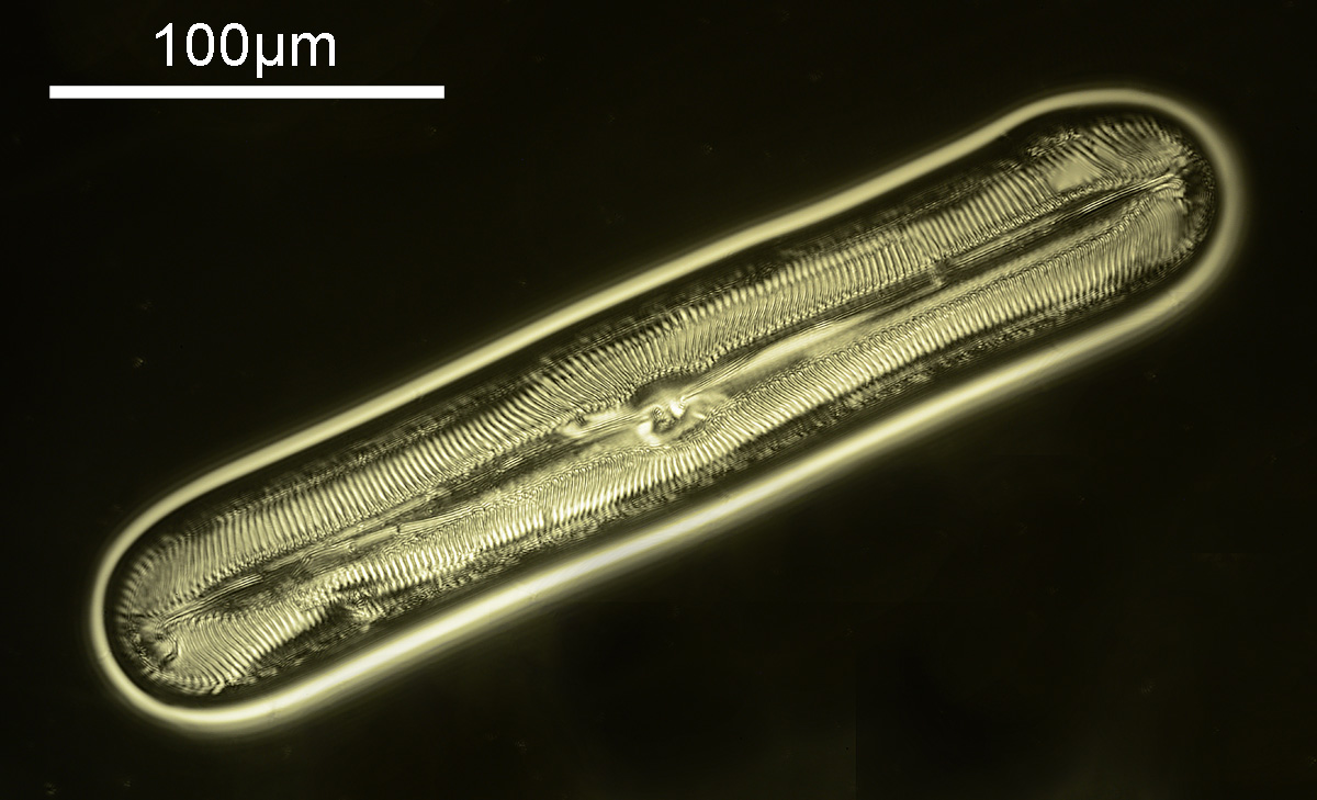
By closing the iris on the condenser right down it does indeed produce a nice darkfield image. By using the quartz condenser and slide, and combining this with a mirror objective it was possible to create a darkfield image in the UVB region at 313nm, something which I hadn’t previously thought possible with the equipment I currently have. For the fellow optics geeks out there, here’s a picture of the objective.
Sometimes it is worth doing experiments just to see what will happen, as the results can be unexpected and open up new possibilities. So, don’t be afraid to experiment. As always thanks for reading, and if you’d like to know more, I can be reached here.
EDIT 3/7/23. Turns out this principle of using a smaller aperture in the condenser with a mirror lens to create a darkfield image has been reported before and is called ‘luminance contrast’. I just spotted mention of it in “Piper, J., & Chmela, G. (2011). Advanced Techniques for Observation and Photomicrography of Subcellular Structures in Diatom Shells. Microscopy Today, 19(1), 10-14. doi:10.1017/S1551929510001203“. They refer back to two other papers for more information, “J Piper, Microscopy Today 15(4) (2007) 26–34” and “J Piper, Int J Light Electron Opt 120(18) (2009) 963–75, doi: 10.1016/j.ijleo.2008.03.032“.