
Short update today, as this is a work in progress. With UV microscopy, conventional glass lenses absorb the short wavelength UV, and become opaque. This means using quartz, fused silica, calcium fluoride, etc for lenses especially when going below 300nm. I have some commercially made UV objectives, but nothing with a low magnification – the lowest magnification I have is a 10x one (which equates to a 16mm focal length on my setup). Having a wider field of view is nicer for looking at big samples, but there is of course a trade off in resolution. This got me wondering if I could make my own objective using a simple fused silica single lens, which is the topic of this post.
To make it I went back to my old friends – Thorlabs – for the components. For those of you who don’t know, Thorlabs is an excellent optics supplier, a sort of Lego for optics geeks, and offer a huge range of components. I’ve written about them before here. For the initial attempt I decided to use a 40mm focal length, 25mm diameter, plano convex fused silica lens, as I had one already. This was mounted in a Thorlabs SM1 tube, and then a RMS to SM1 adapter so it could be screwed into a normal microscope objective port on the microscope. I also fitted another SM1 tube to act as a light baffle to help remove stray light. Here it is on the microscope, pointing at the sample.

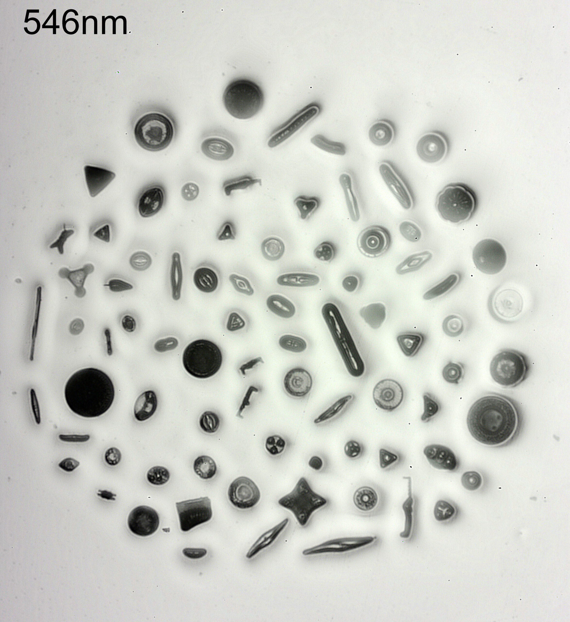
The sample is my fused silica diatom slide for UV work. The diatom arrangement is about 2mm across, and is far too big even for a 10x objective.
Here’s how it looks using visible light (546nm).
And now with 313nm light.
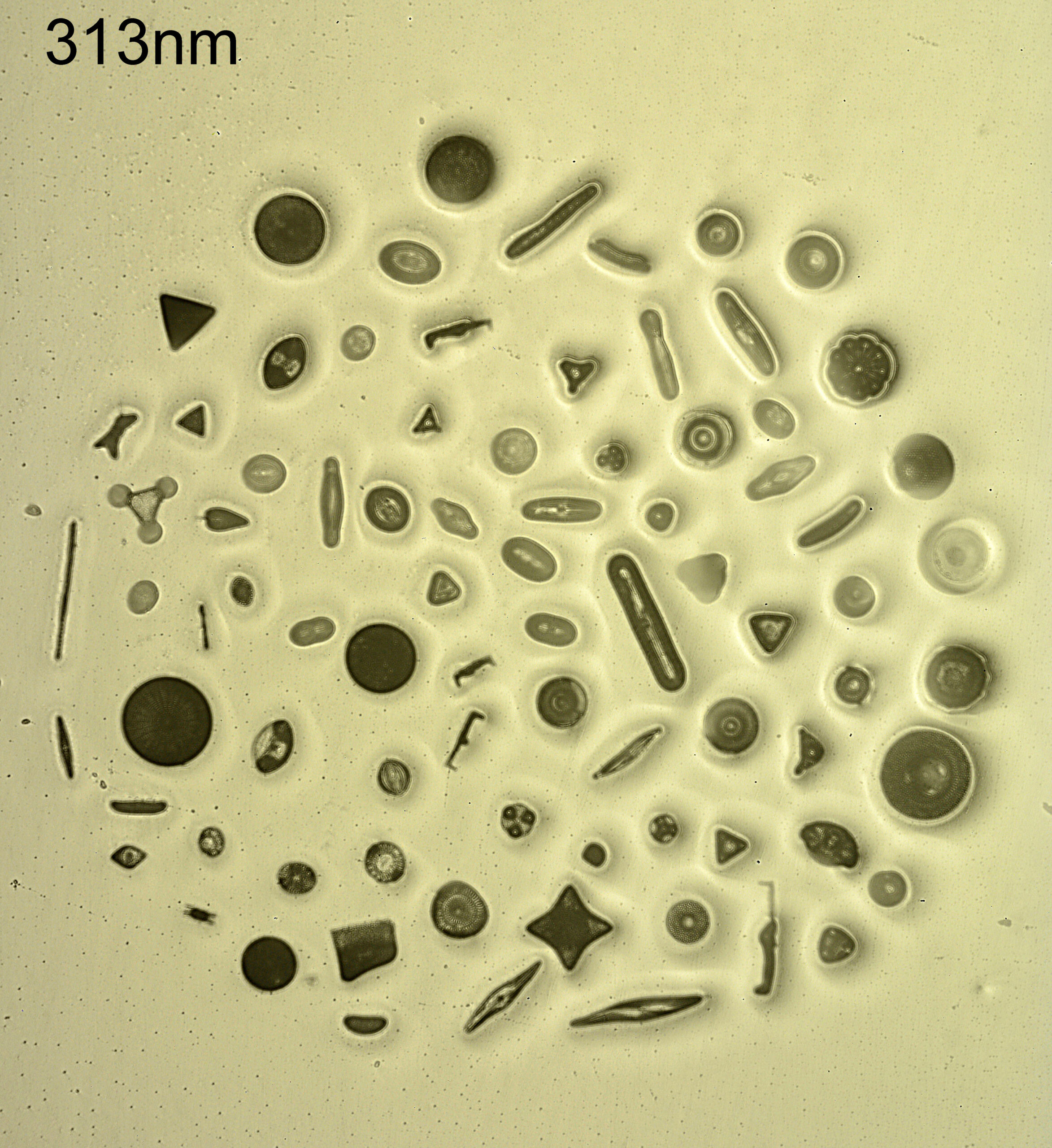
The good thing is it does image the whole of the arrangement, but they are very soft images. Not a huge surprise, this was a single 40mm focal length plano convex lens after all. This was more a proof of concept than anything else. The dirt in the mount it more obvious at 313nm. Again not a massive shock as the shorter wavelength light shows more dust and dirt. Some (but not all) of the diatoms look darker at 313nm than at 546nm, and this is what I have seen with my imaging. There is also a slight change in size of the field of view when changing wavelengths – 546nm and 313nm focus at different places with this lens, so the sample needs moving as the wavelength changes. This is easier to see if I flip between the two images (click on the image for this to work).
Can I make my own objectives using stock components? Yes. Would I want to use a simple plano convex lens for my microscopy? No. Too soft and too low a contrast. I have got a couple of aspheric lenses on order from Thorlabs (one is fused silica the other in glass) to see how they work better – being aspheric I am hoping for a slightly sharper image from them.
As always, thanks for reading, and if you’d like to know more about my work, I can be reached here.