
For those of you that don’t follow my work, I like to use historic optics on my microscope and see what they can do. A few weeks ago I got a Leitz darkground condenser made from quartz instead of glass (which I wrote about here) as I wanted to try it for my UV imaging research. The condenser was screwed into a holder, and so far I have been unable to free it off from that (likely it hasn’t been unscrewed in about a 100 years). As I wanted to use this on my microscope the simplest answer would a cylindrical adapter to fill the space between the condenser and the microscope’s condenser holder. A simple enough task for a machinist or someone with a 3D printer, but I am not a machinist, nor do I have a 3D printer. So I approached a UK company – Flex 3D Printing Ltd – with my rough sketch and they were quickly able to print me up a cylindrical adapter. Today I’d like to share some imaging work done with the condenser now that it can be mounted in the microscope.
First a image of a diatom slide (more on the slide in a minute) using a 10x Nikon Plan Apo NA 0.45 objective and 450nm LED light. The Leitz Quartz darkground condenser was oiled to the underside of the slide (glycerin would have been preferable, but oil was fine in this case).
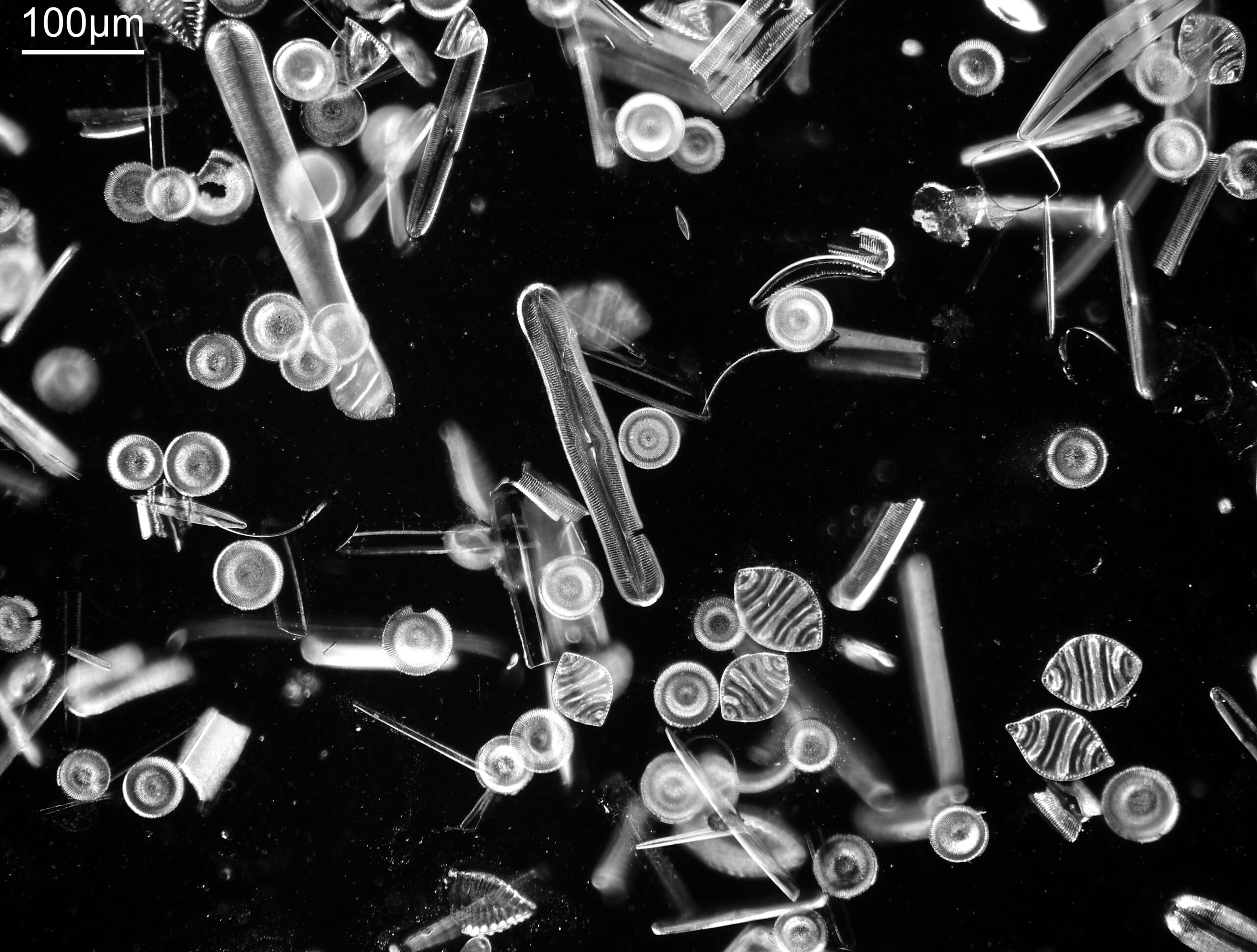
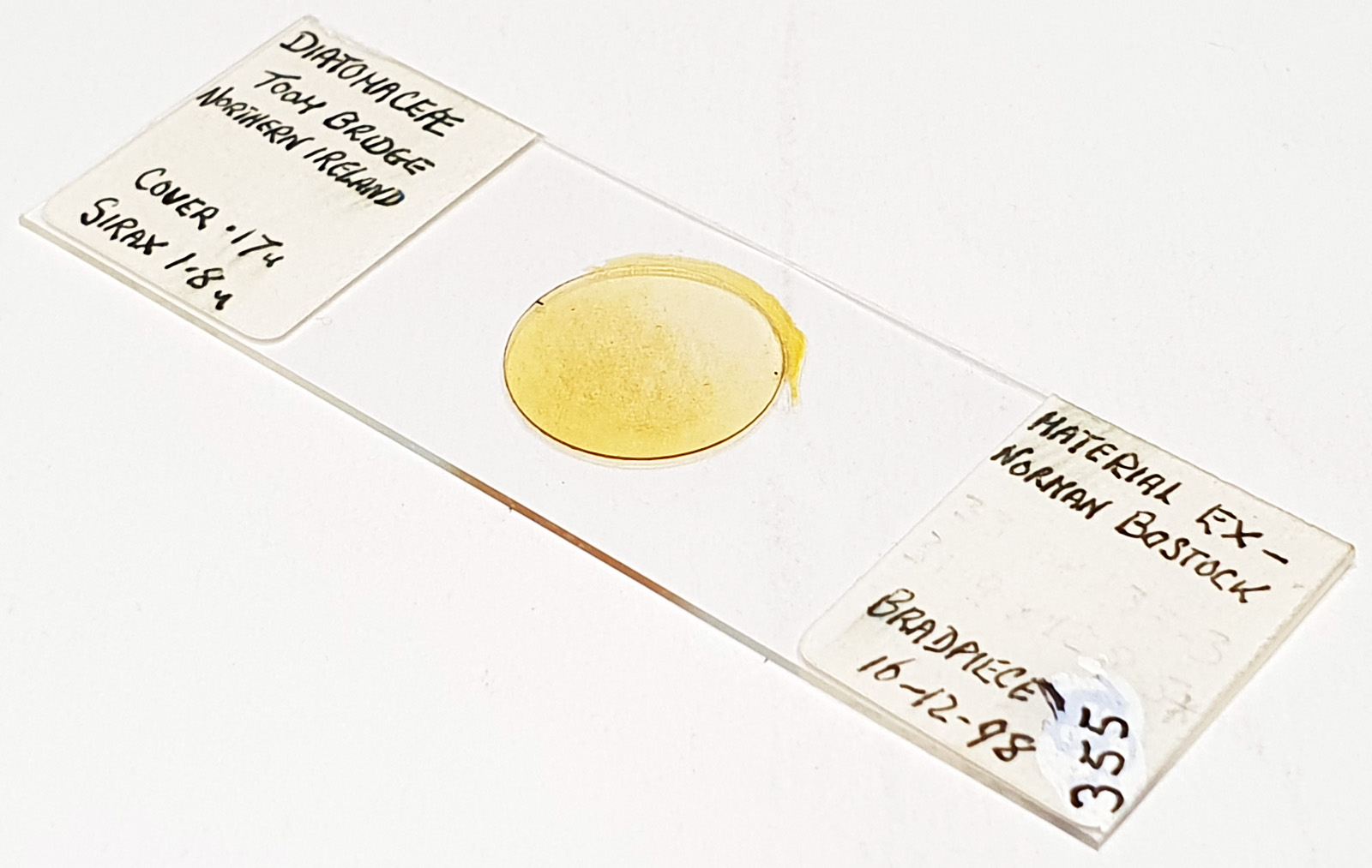
The slide has diatoms from Toome Bridge, Ireland, and shows a good range of species. The long one with rounded ends near the middle of the image is a Pinnularia of some type. This (and the small fragment to the left of it) will be the area I concentrate on for the rest of the images. All the images are single shots, not stacked. Focus was done by eye as a ‘best guess’ using live view on the back of the camera.
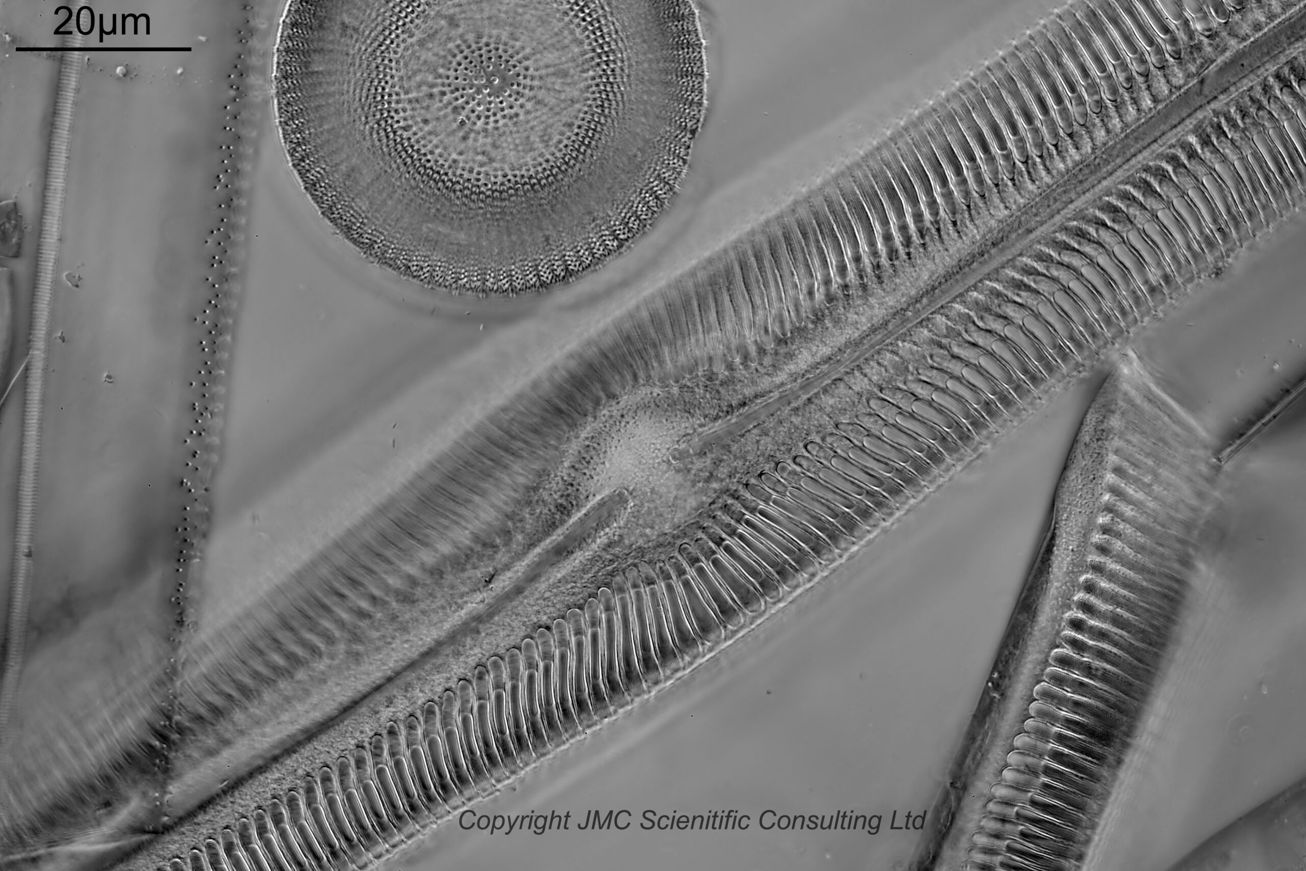
Next is an image using a 40x Leitz Pl Apo NA 1.00 oil immersion objective. For this and subsequent images the orientation has been rotated 90 degrees.
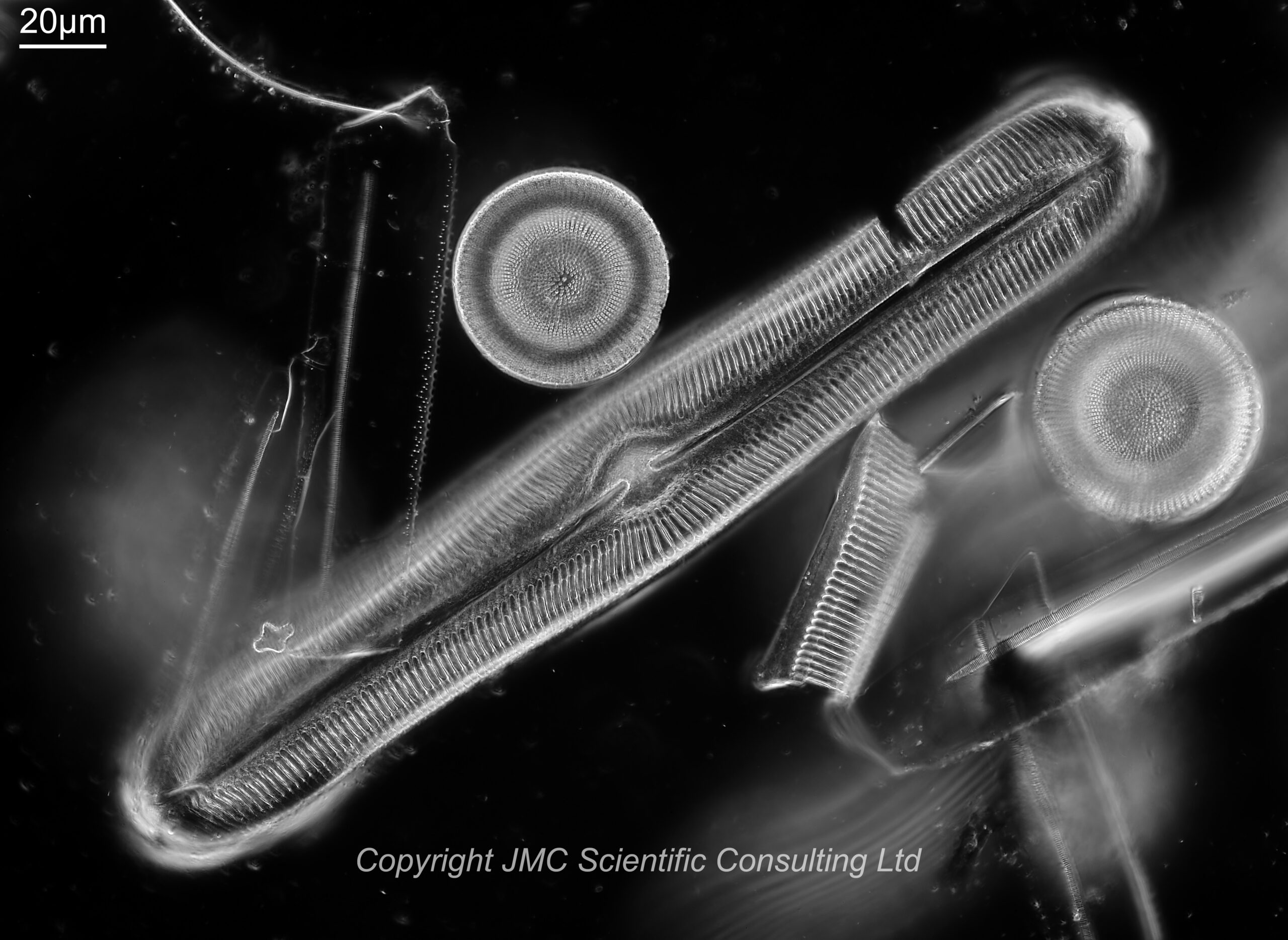
I used 450nm light for this image. Along the centre of the Pinnularia is a raphe line with a small gap in the middle. Perpendicular to this running to the edge of the diatom are striae (these have rounded ends near the middle of the diatom). To the lower right of the main Pinnularia is a fragment of another one. With this objective the striae are easy to see, but the smaller structures within them (called poroids) are not.
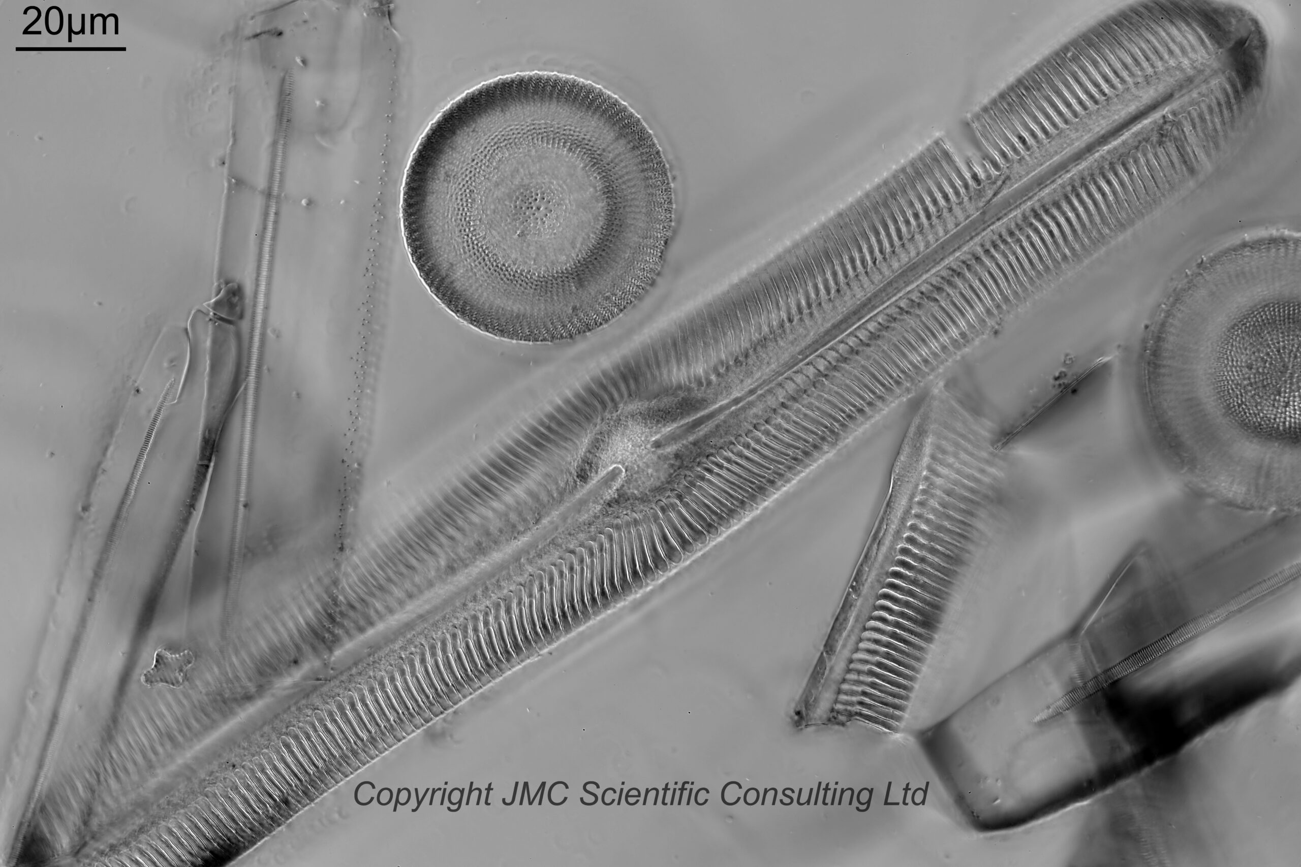
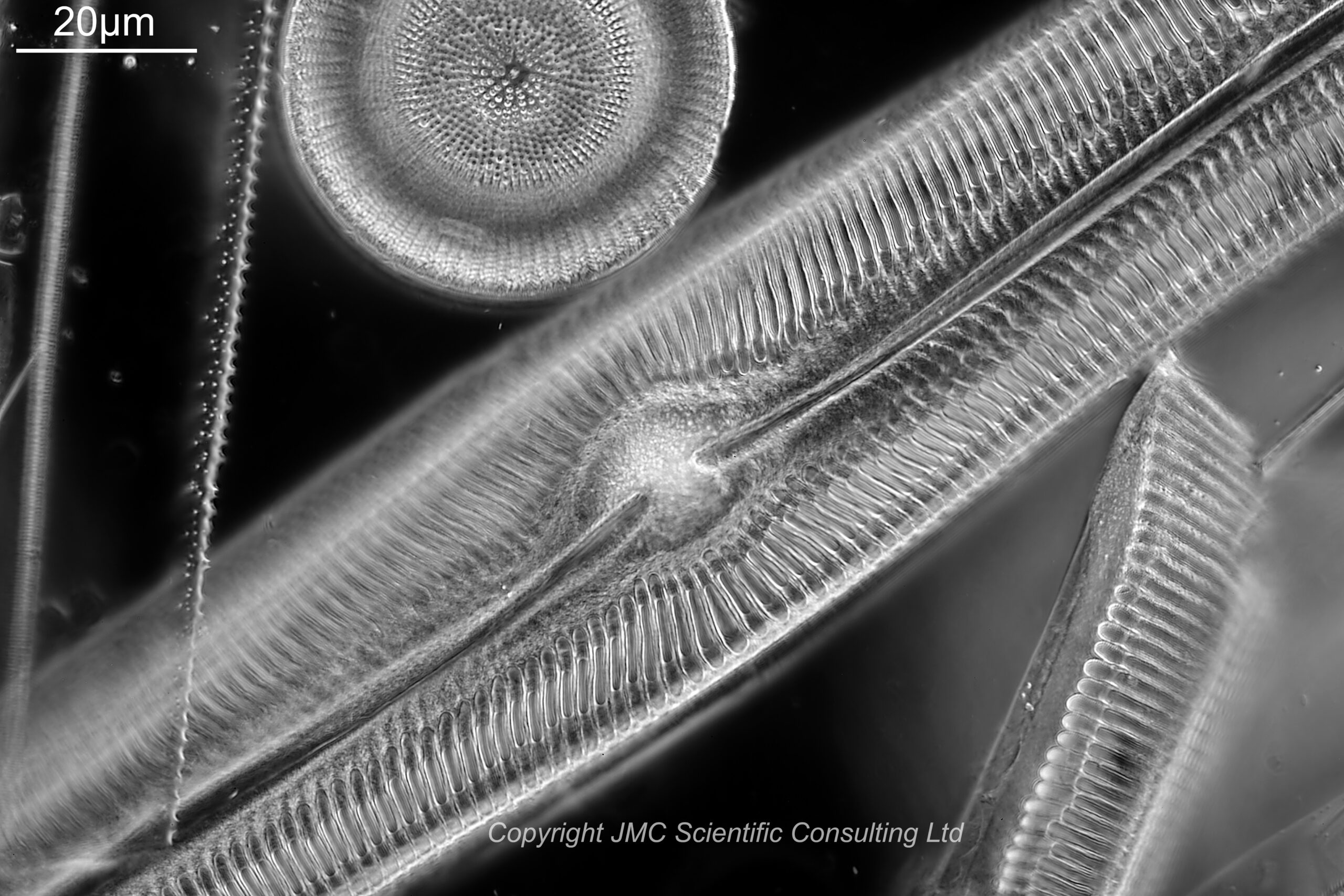
Next up in magnification is the 63x Leitz Pl Apo NA 1.4 objective (used with oil immersion). First using 450nm light.
And then using 365nm UV light.
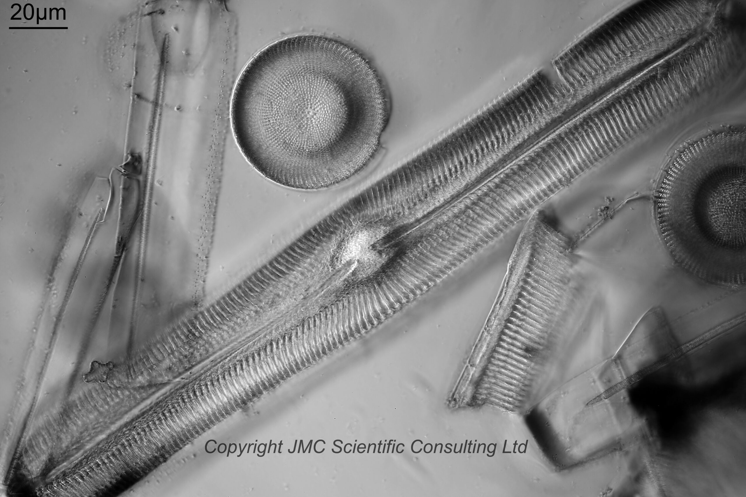
Both images with the 63x objective do not look like darkground images with the black backgrounds. The NA of the objective is now higher than that of the condenser (which is about 1.2) so a true darkground image isn’t made. This type of image is more ‘circular oblique’ lighting, with the light coming in from all around but at a steep angle.
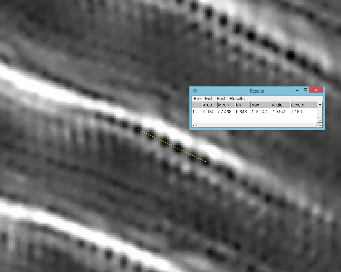
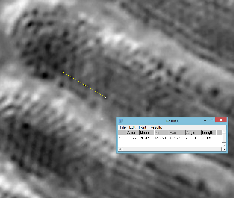
Going in close on part of the image (actually part of the fragment to the lower right), it was now possible to see the poroids within the striae of the Pinnularia and to measure the spacing between them. Below are two screen grabs from ImageJ. First from the 450nm image.
Then with using 365nm UV light.
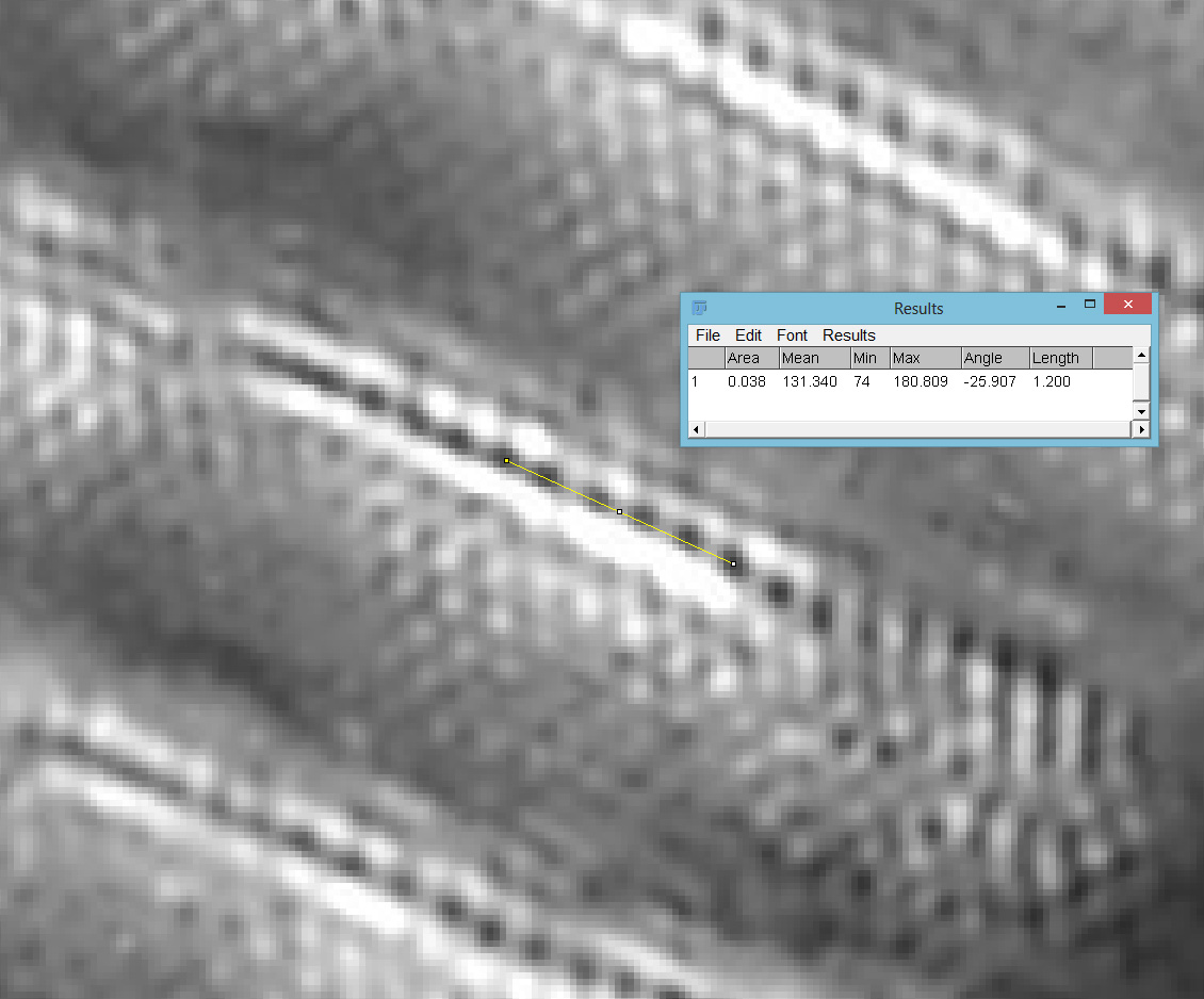
The poroid spacing (centre to centre) was measured as 236nm on the image with 450nm light and 240nm when using 365nm light. Moving from 450nm to 365nm illumination changes the focal position (and therefore magnification) of the objective, so I have to account for that. Given that the measurements of the poroid spacings were only just over 1% different this is within acceptable errors. Resolving poroids on Pinnularia using optical microscopy is a real challenge, so it was great to see this. These features are less than a quarter of a micron apart. A quarter of a micron is the size of a small bacterium, or less than 1/200th the diameter of a human hair.
The 365nm image should be better resolution than the 450nm one (shorter wavelength better resolution). Given my manual focusing I can’t be sure I am actually seeing that, however in the full size images it seems to show better contrast if nothing else.
I did have another objective to try, a Leitz 100x Pl Apo NA 1.32-0.60 with an adjustable iris. Two images were done with this, one with the iris fully open and the others with the iris closed down just enough to make the image darkground. Both were done with 450nm light. First with the iris fully open.
Next with the iris closed down enough to make the image darkground.
Closing the iris reduces the NA of the objective, and as it goes below that of the condenser, the image goes from ‘circular oblique’ lighting to darkground.
I also did a screen shot of the Pinnularia fragment (although a slightly different part of it this time – focusing on the live view of the camera is not always ideal). This was done with the iris fully open.
With the iris fully open and with 450nm light, the poroids were visible (and measured spacing was 237nm with this objective), but with a slightly softer image than the 63x objective. This would be expected, as although the 100x objective offers more magnification, it’s NA when the iris is wide open is lower than that of the 63x (1.32 vs 1.40). However given the higher magnification, there are more pixels to play with and the image is smoother. I am not showing a magnification of the image with the iris closed down. Although the image was darkground, closing the iris reduced the resolution and the poroids were not longer visible. Swings and roundabouts……
So, overall the condenser performed well and allowed imaging of poroids in a Pinnularia diatom with spacings of around 250nm, and allowed the use of UV light (365nm) as well as visible light.
Before I wrap this up, some info about the slide and the adapter. The slide was by Neville Bradpiece, and is a strew from Toome Bridge, Northern Ireland. The diatoms are mounted in Sirax which is a high refractive index mountant.
The adapter was made from PA12 Nylon. The images below show it by itself and mounted on the condenser.

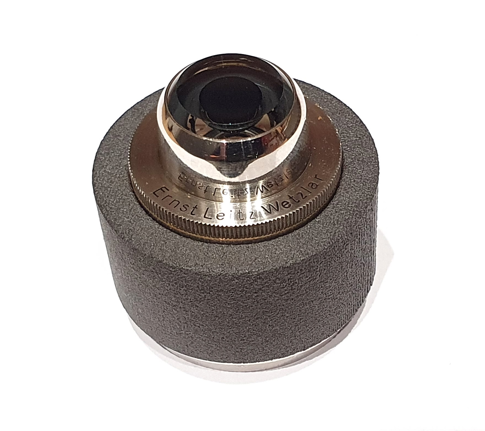
The condenser was a friction fit on the inside, although in future I will add a little double sided tape for extra security (I do not want the condenser falling out the bottom of the adapter and crashing into the field lens on the microscope, especially when that is a fused silica one).
Big shout out to Flex 3D Printing Ltd. I’m always happy to support UK businesses and without folks like these my work would be a lot more difficult.
As always, thanks for reading, and if you’d like to know more about my work I can be reached here.