I’ve been busy with work, but last night took a break to look at a microscope slide which arrived recently. It was described as a ‘Polycistina’ slide from Barbados and it had an unusual feature on it.
Here’s a couple of pictures of the slide taken using different iris settings on the condenser.
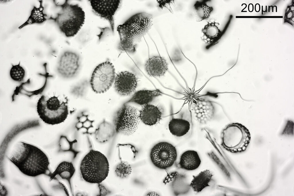
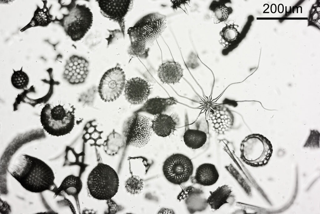
The unusual feature is the one with tendrils, and it has been mentioned that this could be a plant trichome. In the images above, there were two settings used for the condenser iris – the first one was open to NA 0.4, and the second one it was closed down to about 0.2. Everything else was the same between the two images (Nikon d800 monochrome camera, 10x Olympus UVFL NA 0.4 objective, white LED light) although exposure time was increased for the smaller iris aperture. This shows the effect that iris aperture setting can have on depth of field in an image. Closing down the aperture too far though can be a bad thing due to diffraction starting to occur, and also a shallow depth of field helps emphasize the subject. As with many things in life choosing what looks right is a balancing act.
The sample is quite thick, and as the stage was moved up and down different parts of the image came in and out of focus. This can be seen in the video below where the stage was gradually moved up.
It was actually quite interesting to view the slide using a low magnification (4x) objective. The sample was so thick that slight movement of my eyes when looking at it through the eyepieces gave a ‘3D’ image as the features moved differently depending on their depth in the slide.
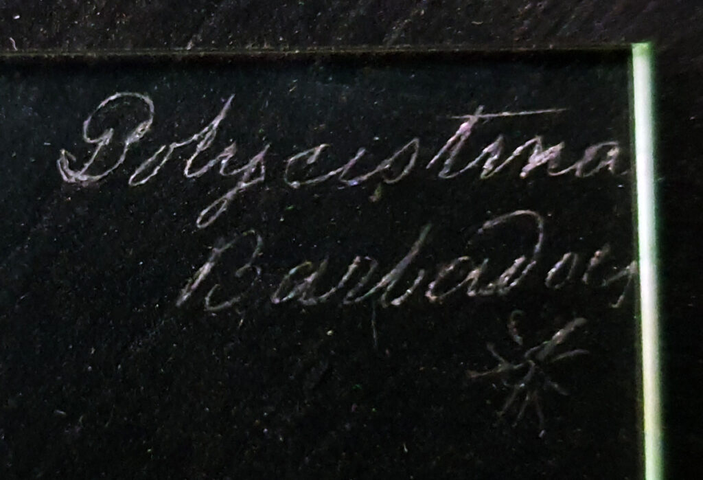
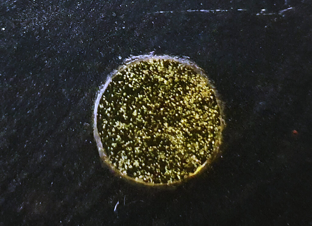
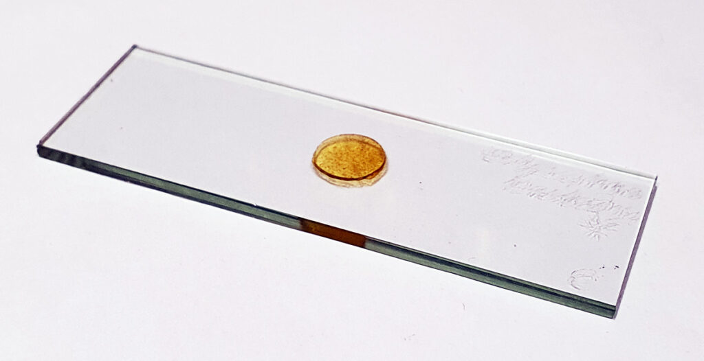
The slide itself is interesting and the maker even engraved the shape of the unusual feature onto the slide. Unfortunately there is no makers name. The slide itself is very thick.
Microscopy continues to fascinate me, providing a window in to the wonderful world of the tiny and the amazing structures that nature can produce. As always, thanks for reading, and if you’d like to know more about this or any other aspect of my work I can be reached here.
