
I’m a huge fan of imaging diatoms on my microscope as they provide such interesting, challenging and varied structures as well as being beautiful to look at. As said on the diatoms.org website, diatoms are ‘algae that live in houses made of glass’. The silica (glass) structures that they produce are what I, and many others, enjoy imaging. Today I’ll share a few of the ones I have imaged recently on my microscope. I tend to use 450nm LED light for most of my visible light work, as the shorter wavelength gives and improvement in resolution for a given setup (see Abbe equation for resolution). In addition to showing the diatoms I’ll also share the slides that they came from. Slide making is an amazing art in itself and the makers deserve to be recognized for their talent. Note I have reduce the resolution of the images to make them easier to share here. Most are actually much higher resolution than I can share on this site.
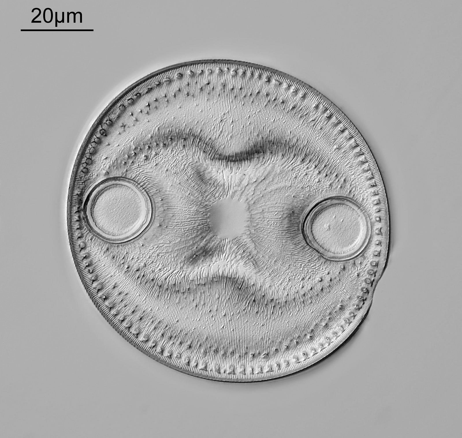
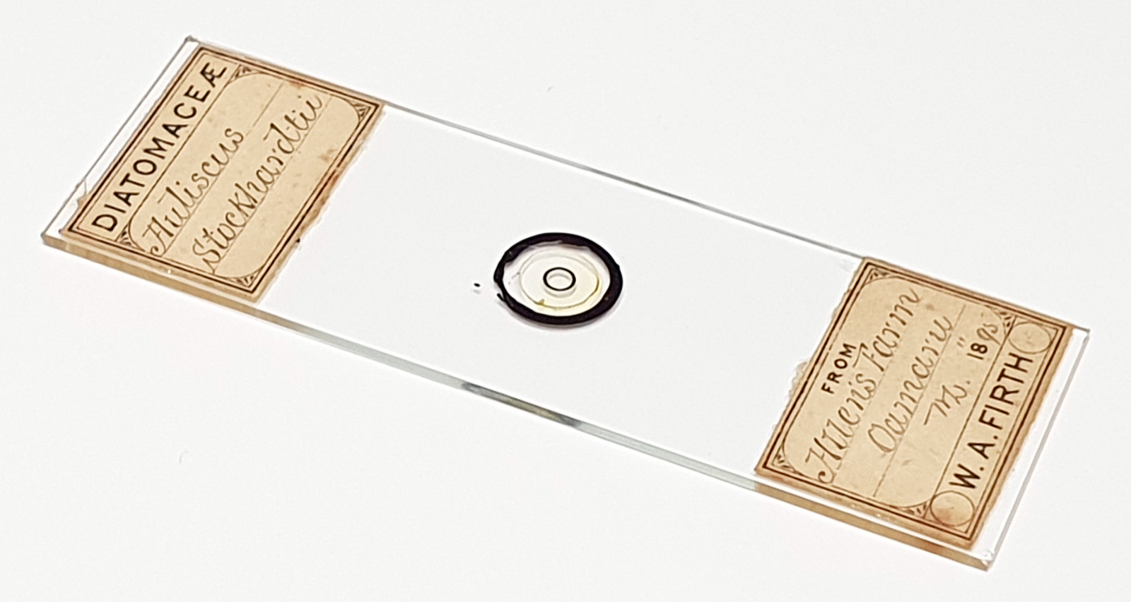
First is Auliscus stockhardtii by WA Firth.
And the slide.
This one comes from Allen’s Farm in Oamaru, New Zealand. Not only is the structure amazing, but is has survived for over 30 million years before being collected and mounted.
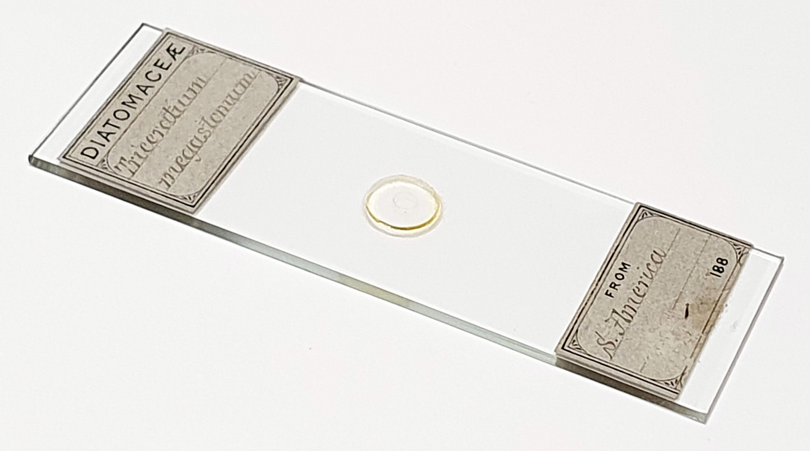
Next is Triceratium megastomum again by WA Firth. This one is from South America.
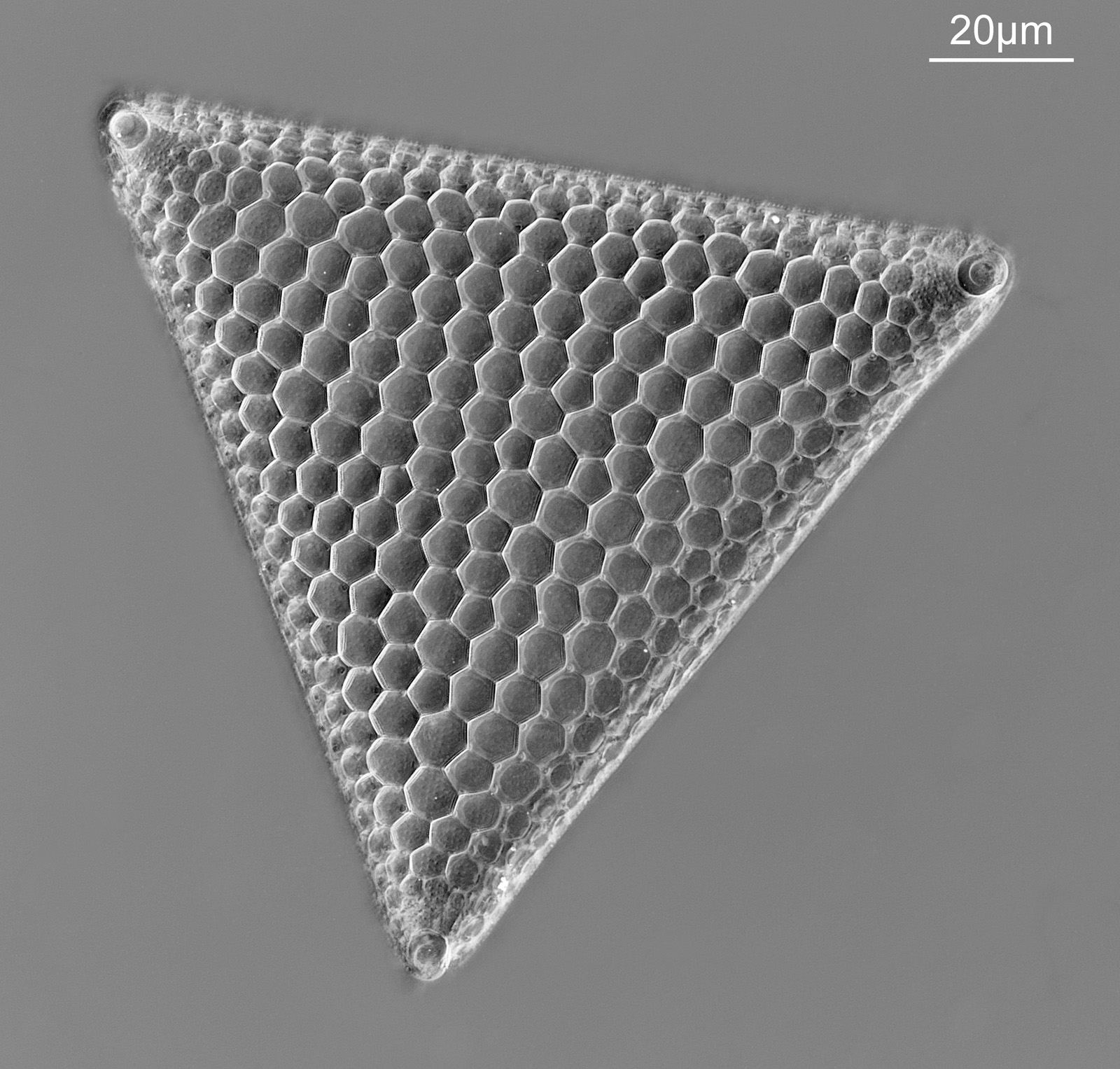
This was originally a bright field image, but I have inverted it to make the photo above. Here’s the slide.
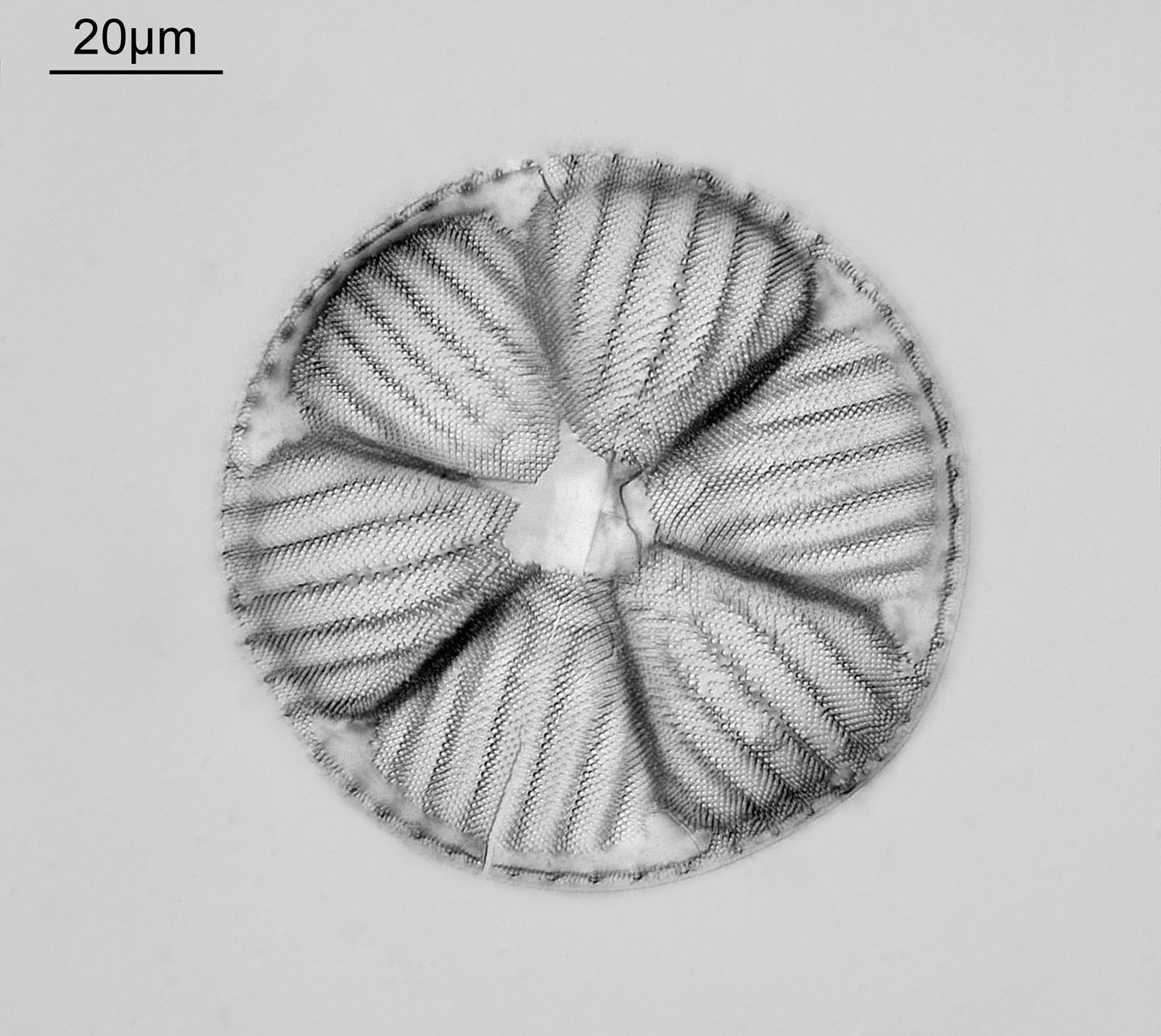
Up next is what I think is an Actinoptychus grovei from a strew slide by Klaus Kemp.
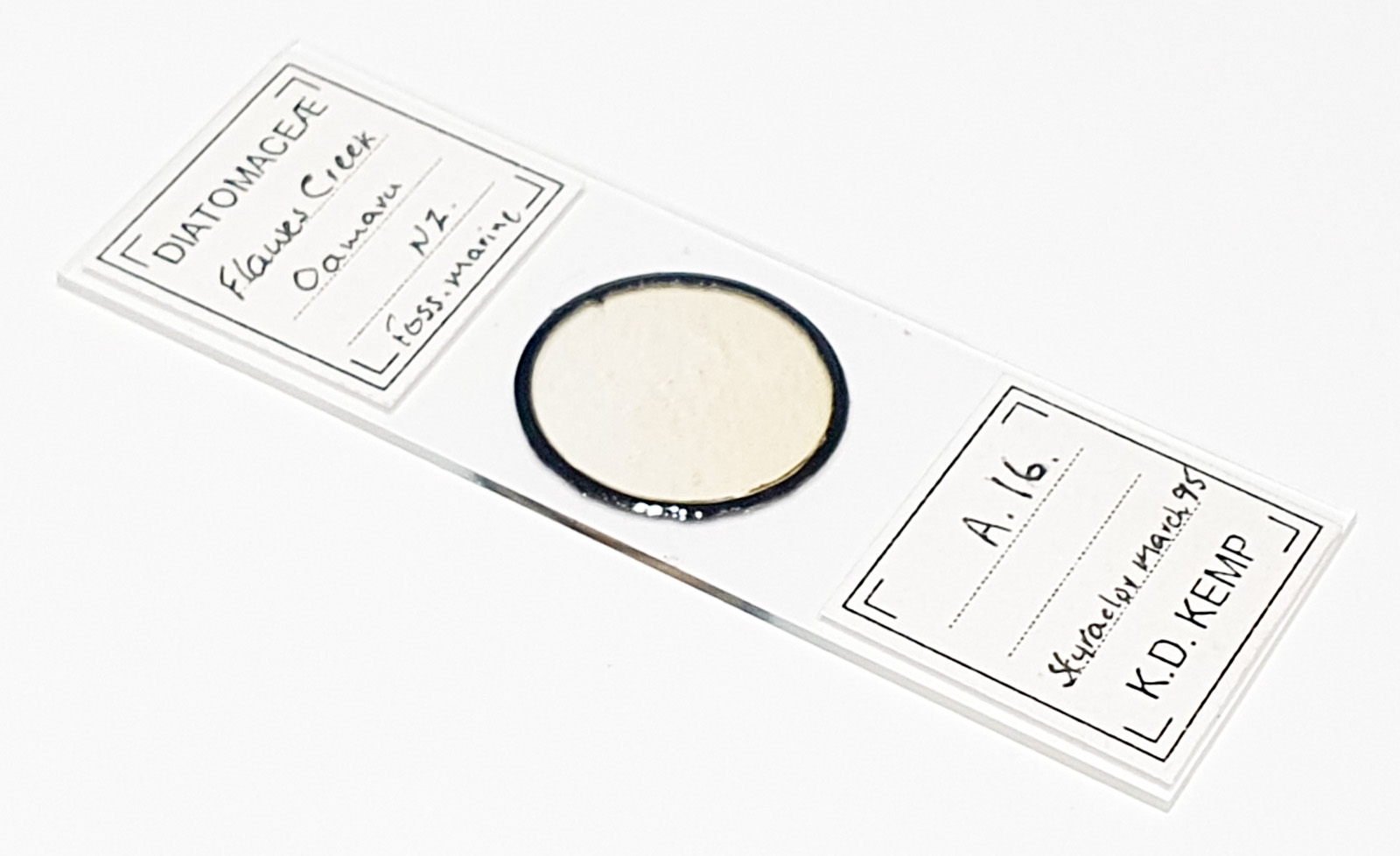
Here’s the slide.
Strew slides offer a lot of different material to image. Some of it is fragments, however there are often some complete specimens as well. It is a bit more of a lottery though – you may get lucky or you may not. My experience has generally been good when sticking to older slides by well known makers. This one again was a fossil one from Oamaru (Flawes Creek) so will be around 30 million years old.
This is from another Klaus Kemp strew slide (from Klamath Falls, Oregon), and I don’t know the name of it unfortunately.
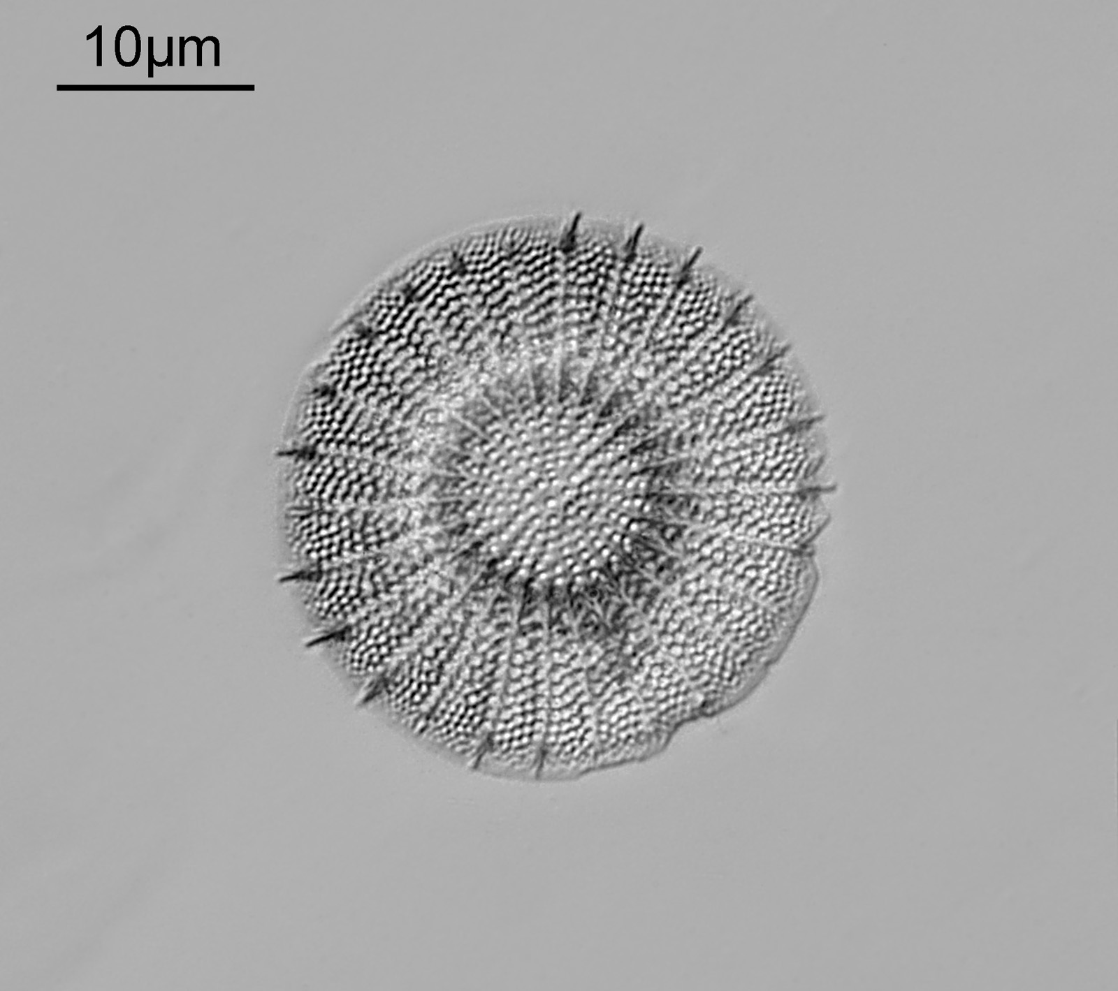
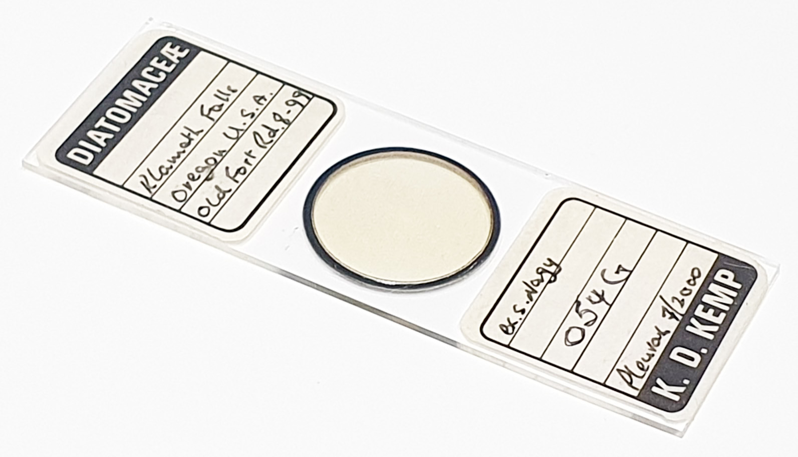
This one was tiny, and I nearly missed it when looking over the slide. And the slide.
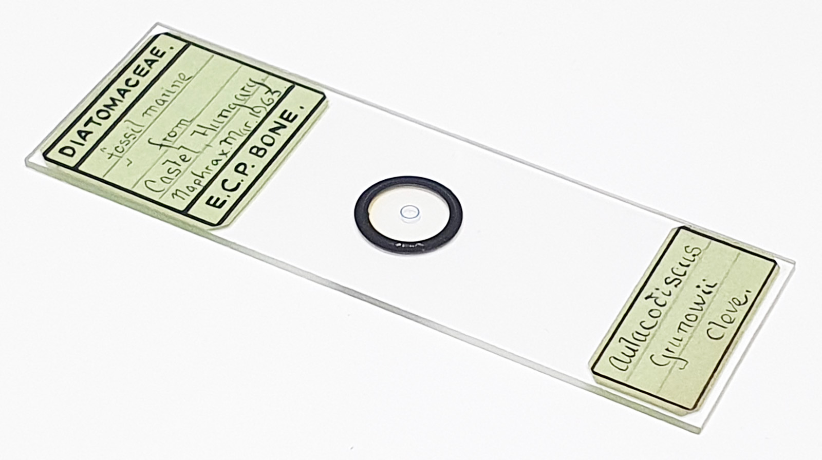
Final one for today. Aulacodiscus grunowii Cleve by ECP Bone.
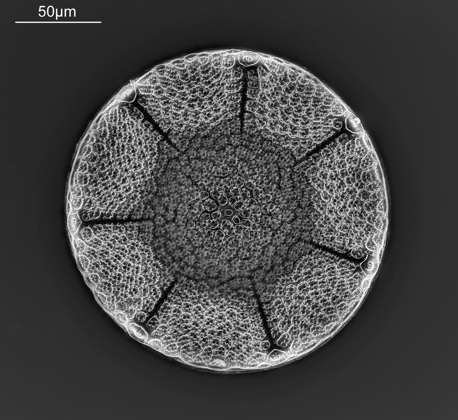
The image was captured as bright field, but I have inverted it as I like the look of the inverted image. Another fossil sample, this time from Castel Hungary. Here’s the slide.
Microscopy allows us to see the beauty in these tine things which to the naked eye look just like specs of dust. if we can even see them at all. I use a relatively basic microscope – an Olympus BHB which I’ve modfiied slightly to allow me to use it for UV imaging as well – but the key is in setting your kit up correctly, using the right lighting and decent objectives. ‘Decent’ does not necessarily mean new, and I typically use Leitz objectives from around the 1970s for a lot of my visible light work.
Anyway, I hope you have enjoyed these, and I will share more in the future as I have a few slides to work through. Thanks for reading and if you’d like to know more about my work I can be reached here.