I’ve had a bit of a catch up this weekend and looked at some slides that have arrived recently. They are both pretty and interesting (for different reasons), so I thought I would post them here. All have been reduced in size for sharing.
First is a Pinnularia slide by Arthur Cole.
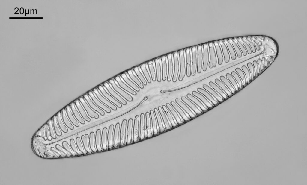
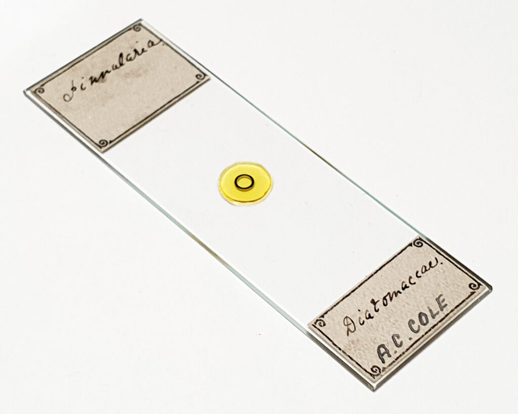
There was a single example of the diatom on this slide. The mount was amazingly clear and needed very little cleaning up. Imaging was simple bright field, using 450nm light. Date for this is likely the late 1800s.
Next we have a Triceratium scitulum slide by Thomas Russell of London.
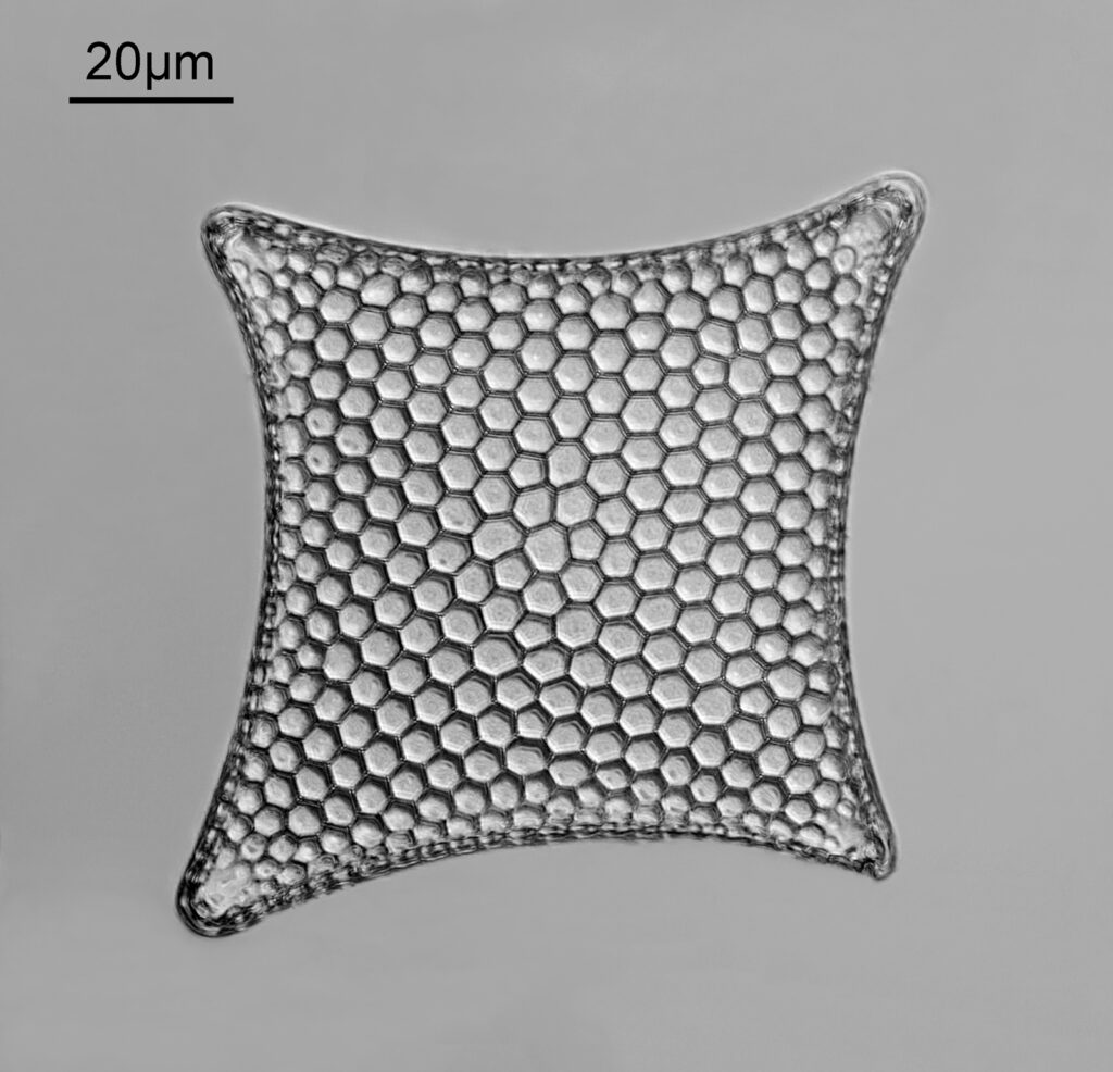
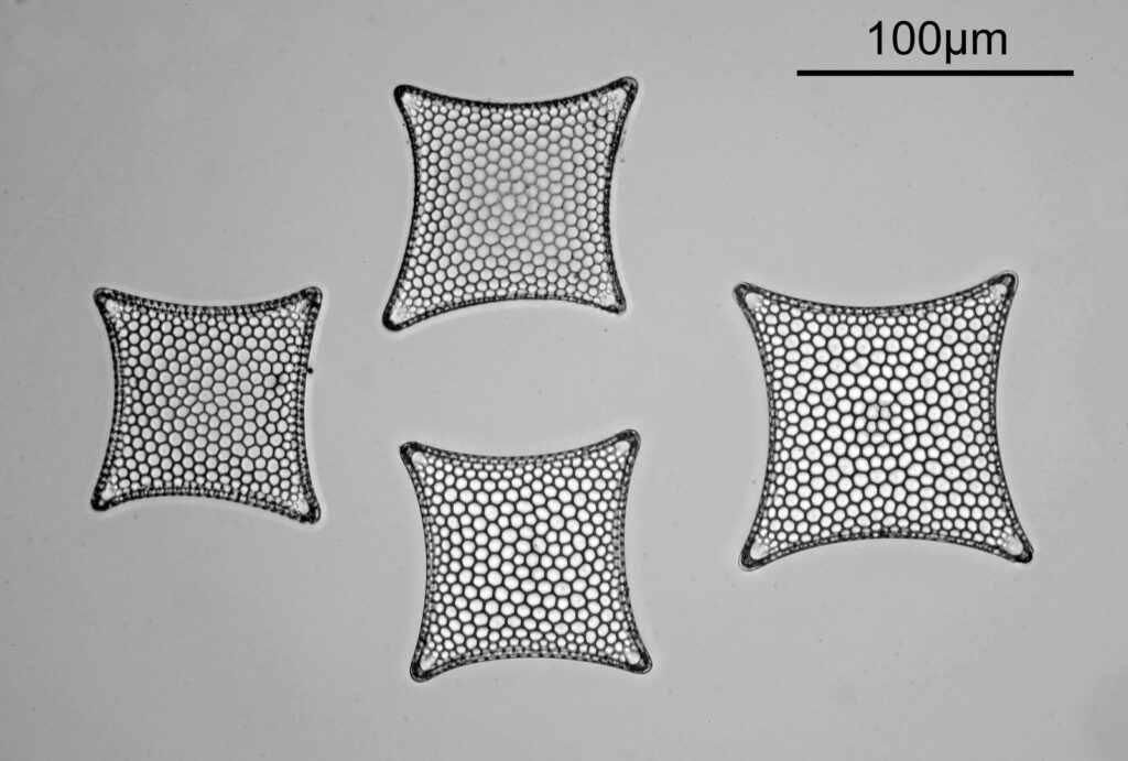
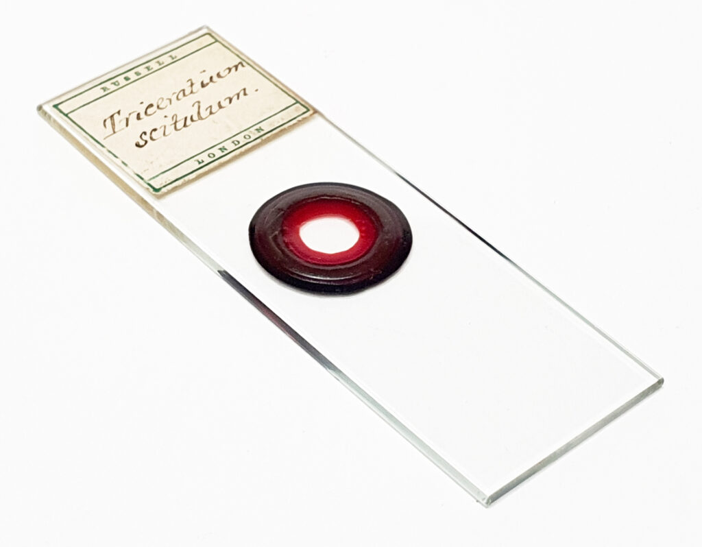
A nice arrangement of 4 diatoms. Again the mount was very clear and needed very little cleaning up. Imaging was brightfield with 450nm light. Likely date for this one, late 1800s.
I returned to this slide the next day and took a picture of one of the diatoms using the Watson Holoscopic condenser to give a dark ground image.
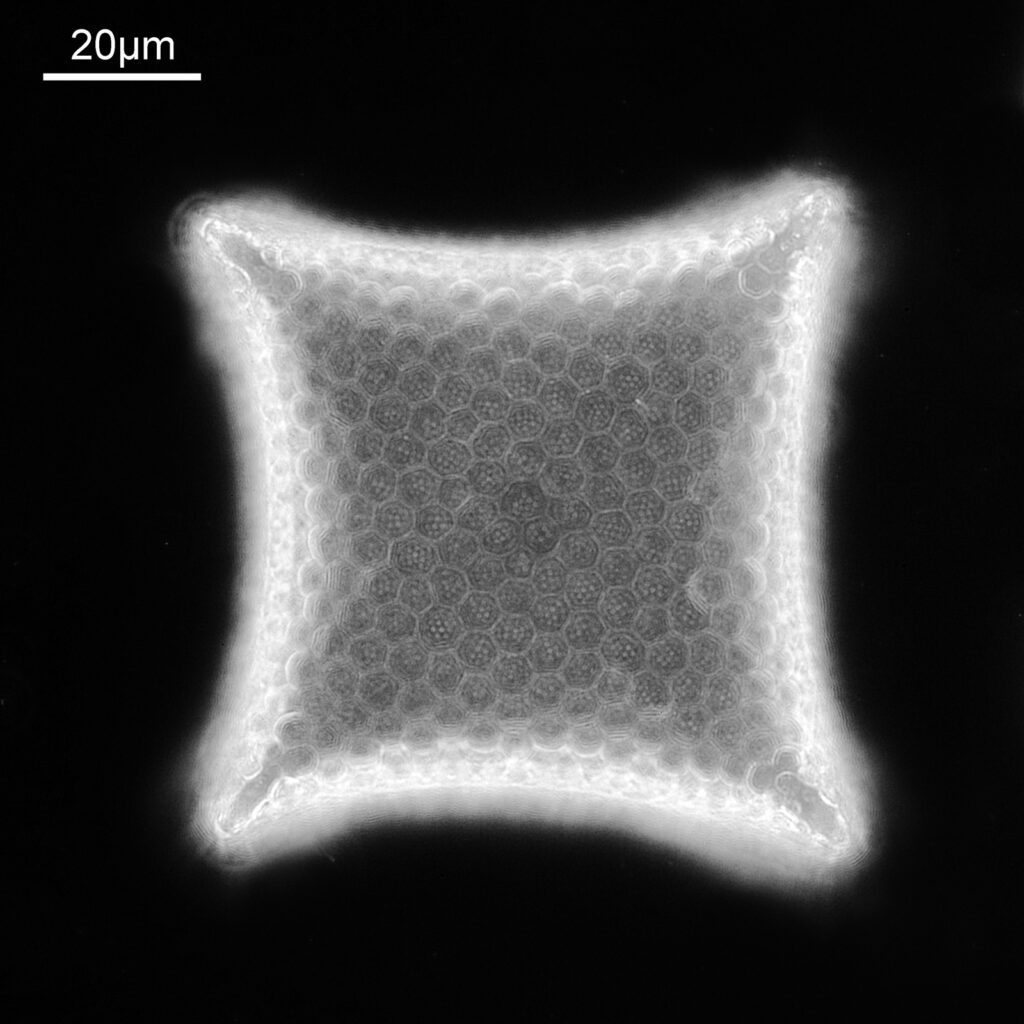
Using the Holoscopic condenser has brought out the dot features present within the polygonal structures which were barely visible in the bright field image. The ‘dots’ look to be about 500nm across.
Now an arrangement of Actinocyclus by Eduard Thum, Leipzig.
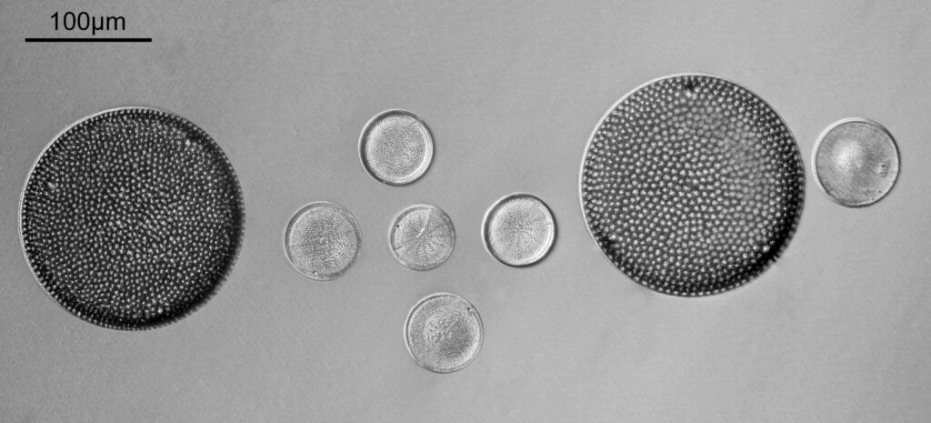
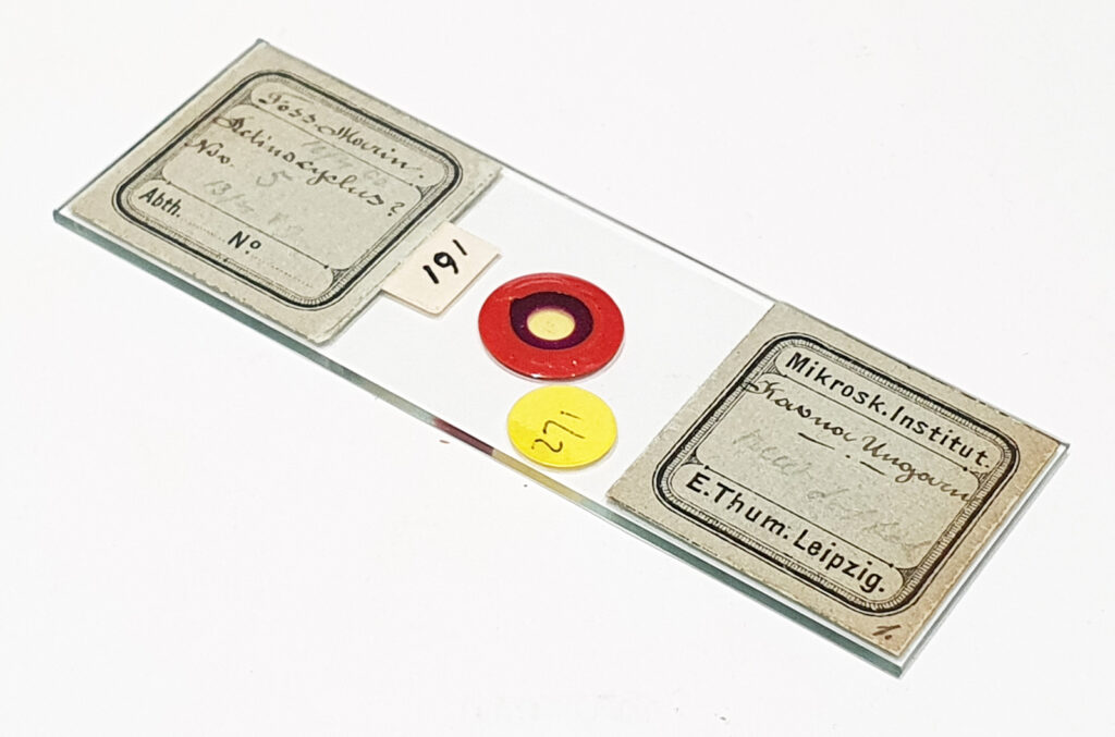
A typical Thum arrangement using two large circular diatoms to mark where the arrangement is. Imaging was oblique brightfield with 450nm light. Likely date for this one, again late 1800s.
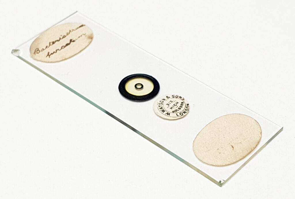
Last but not least is a Bacteriastum furcatum slide by Watson.
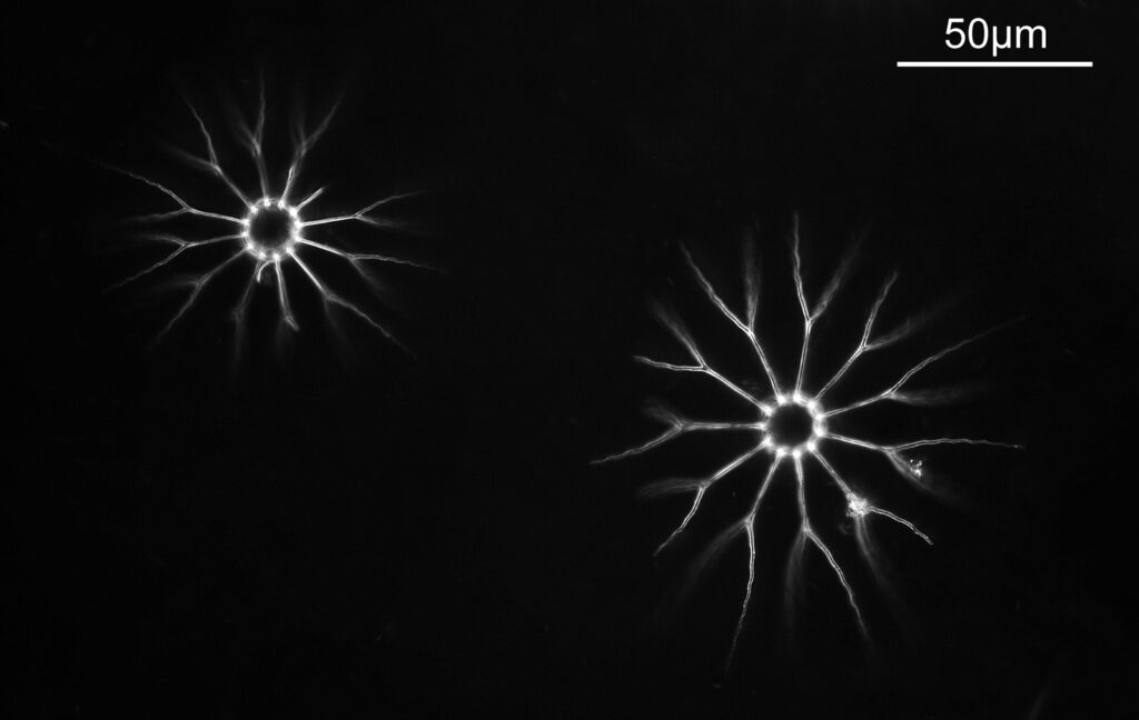
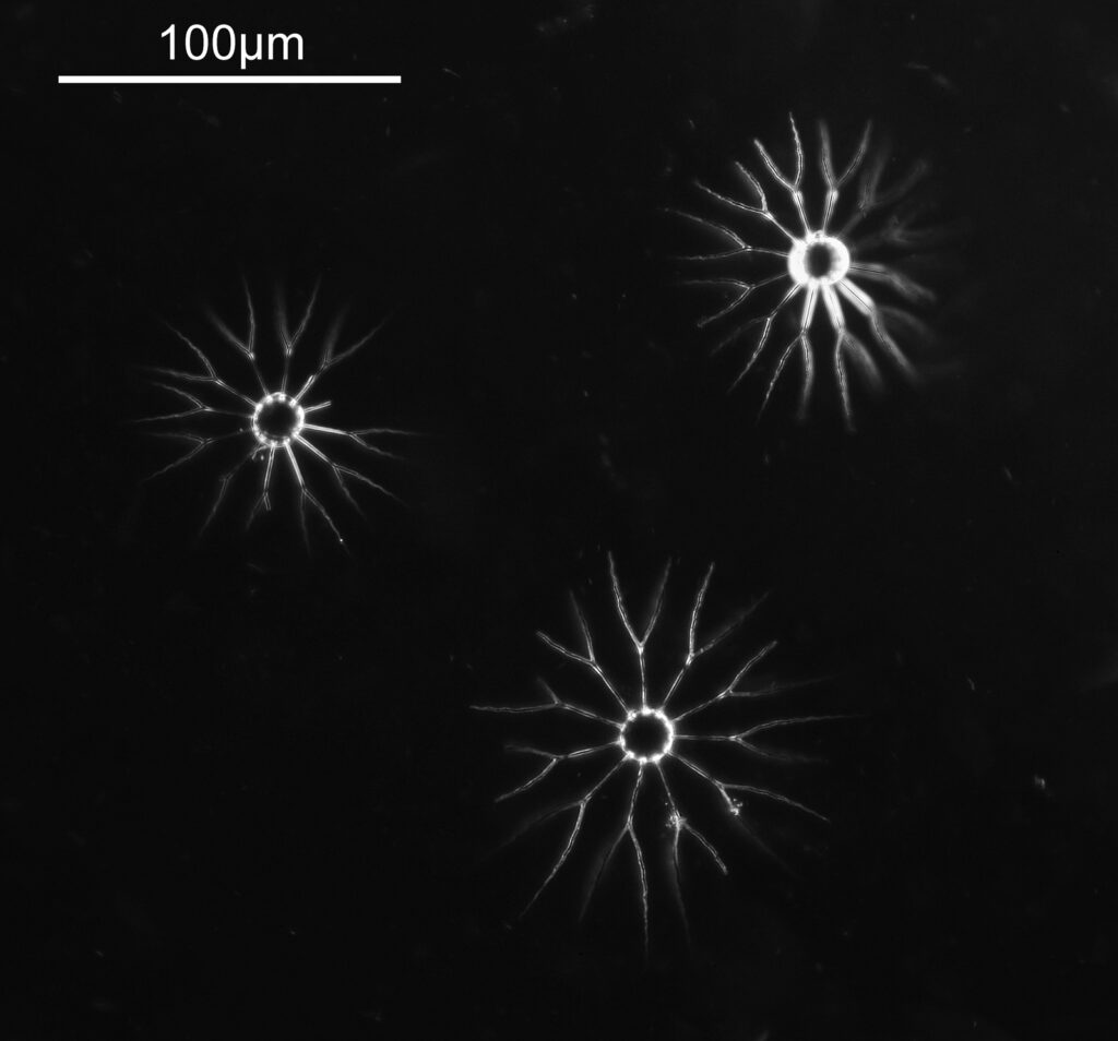
Imaged using 450nm light and a Watson Holoscopic condenser (discussed here) to give the dark ground image. Single images, no stacking.
There are always lots of these interesting old diatom slides available on places like Ebay and in my experience they offer great value for money for those wanting to image slides, so don’t discount them because they are old. As always, thanks for reading and if you’d like to know more about this or other aspects of my work, I can be reached here.
