At this time of year I try and do a bit of a reflection on what I’ve done over the last 12 months and what’s ahead for the coming year.
My dermatology work has carried on apace this year, both with existing clients and some new ones. I try and publish something each year, and 2024 saw two papers come out in a special edition of the International Journal of Cosmetic Science. These were on the use of In-vivo confocal Raman spectroscopy for looking at skin, a technique I was heavily involved with the development of. One looked at the development of the technique and the other how it was used in combination with a range of other skin measurement techniques to better understand how dry skin impact skin properties. This was a study I designed and ran back in 2006-7 but it was never published at the time. The techniques are still relevant today and I thought it was a good time to get this out there (take a look in my publications section for the two papers, they are free access so don’t require a subscription to download).
I’ve also been working with a couple of clients to characterize the spectral responses of their cameras. With this I measure the response of the camera between 280nm and 800nm, so can get an idea of how they behave in the UV, visible and IR regions, which is especially useful for people working outside of the visible spectrum. I love this type of research, and it is great to be involved with real imaging and measurement science.
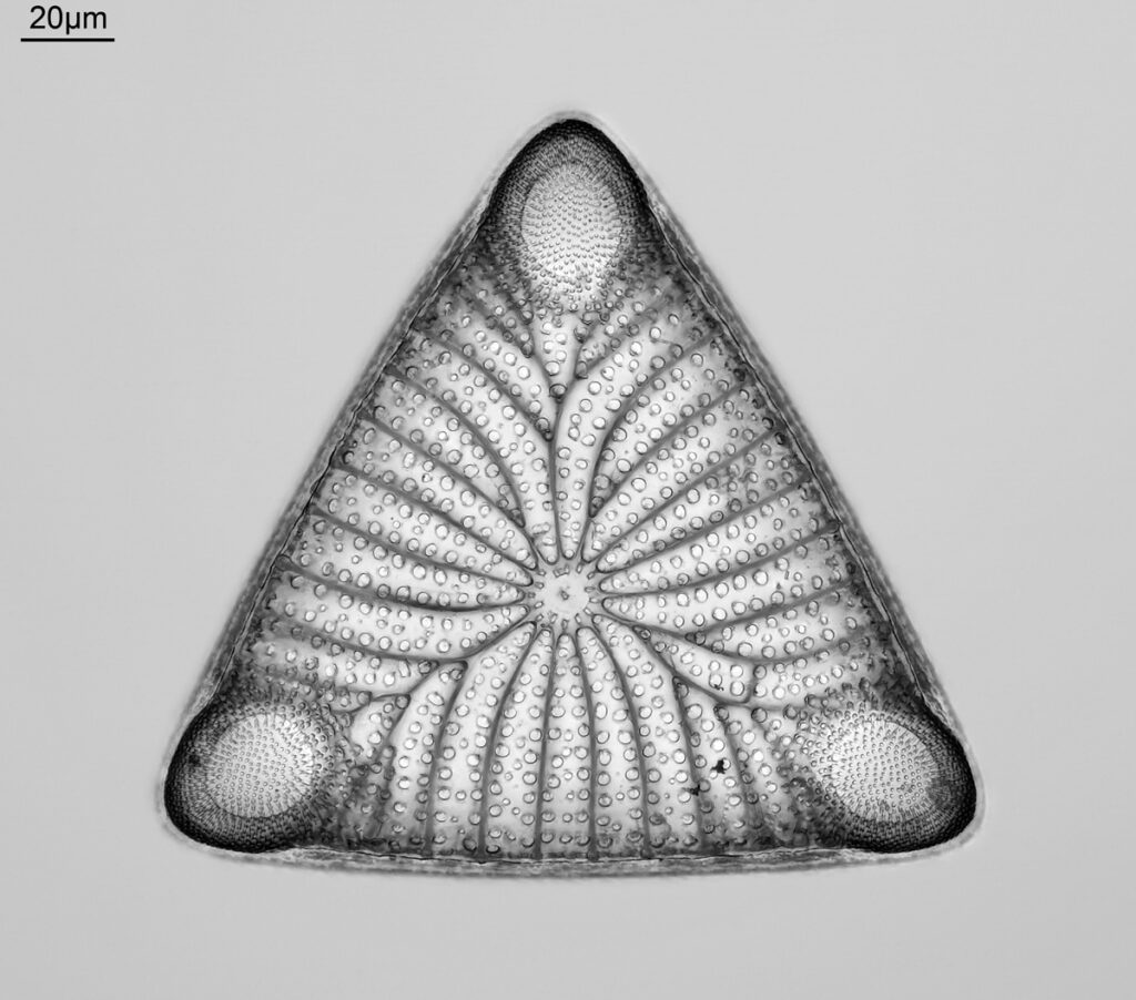
My microscopy work has evolved somewhat, and I set up a new website to share my diatom images. This has the inventive name of Diatom Imaging and here’s the link (https://diatomimaging.com/). On that site I share high resolution optical microscope images from the range of slides I have, along with details on how they were taken and the slides themselves, and is a sort of on-line museum. Many of the diatoms are quite rare, and some I haven’t been able to find good images online, which is one of the reasons I wanted to set this up. An example image of one of the images is given below (this one won an award with the Quekett Microscopical Club).

Although the website has only been active for about 6 months, I have over a 1000 images on it already, and I am adding to it as and when I get the time to image new slides. I’ve already had people reach out to me to ask me to use images, and I hope in the future this will become a significant scientific and artistic resource.
So what does 2025 bring? More of the dermatology day work of course. I have been asked to give a key note talk at the 2025 Society of Cosmetic Scientists annual meeting, which will be a great opportunity to fly the flag for how skin measurements and imaging can be used to improve consumer lives and better understand dry skin.
On the microscopy/imaging side, I will continue to build and add to my Diatom Imaging site. I’m always on the look out for interesting slides to image, so feel free to contact me if you have collections that you are wanting imaging. I also need to return to my UV microscopy work, and do some work work for imaging with light below 300nm. In 2023 I published an article where I showed a new design for a darkground microscope condenser. That was a work in progress, and it would be great to get some time (and money) to finish that off. It needs some investment to buy a new optical lens, so I hope to be able to return to that one in the new year.
As always thanks for reading, have a great Christmas and New Year and all the best for 2025.
